Understanding Spinal Cord Fractures: Anatomy, Causes, and Management
A spinal cord fracture is a serious injury affecting the vertebrae and potentially the spinal cord itself, often resulting in partial or complete loss of neurological function. These fractures can occur in any section of the spine, including the cervical, thoracic, or lumbar regions, with cervical fractures typically posing the greatest risk of severe paralysis or life-threatening complications. The spinal cord is encased within the vertebral column, which normally protects it from trauma. However, high-energy impacts such as falls, motor vehicle accidents, sports injuries, or penetrating trauma can compromise the vertebrae and cause fractures that impinge upon or sever the spinal cord.
Spinal cord fractures are classified based on the type of vertebral disruption. Compression fractures involve the collapse of the vertebral body, often due to osteoporosis or trauma. Burst fractures result from high-impact compression, scattering bone fragments and increasing the risk of spinal cord injury. Flexion-distraction and fracture-dislocation injuries indicate severe instability and often accompany ligamentous damage. The precise mechanism of injury affects both the neurological outcome and the required intervention.
Symptoms of spinal cord fractures range from localized pain, tenderness, and restricted mobility to neurological deficits such as numbness, tingling, weakness, or paralysis below the level of injury. Immediate medical evaluation is critical to prevent further cord damage. Diagnostic imaging, including X-rays, CT scans, and MRI, is essential to assess vertebral alignment, fracture type, cord compression, and associated ligamentous or soft tissue injury.
Treatment strategies depend on fracture severity, spinal stability, and neurological involvement. Conservative management may include immobilization with braces or traction, alongside pain control and physical therapy. Surgical intervention is often necessary for unstable fractures or progressive neurological deficits, aiming to decompress the spinal cord, stabilize the vertebral column, and restore alignment. Postoperative rehabilitation focuses on restoring mobility, strength, and functional independence while preventing secondary complications such as pressure ulcers, muscle atrophy, or respiratory issues.
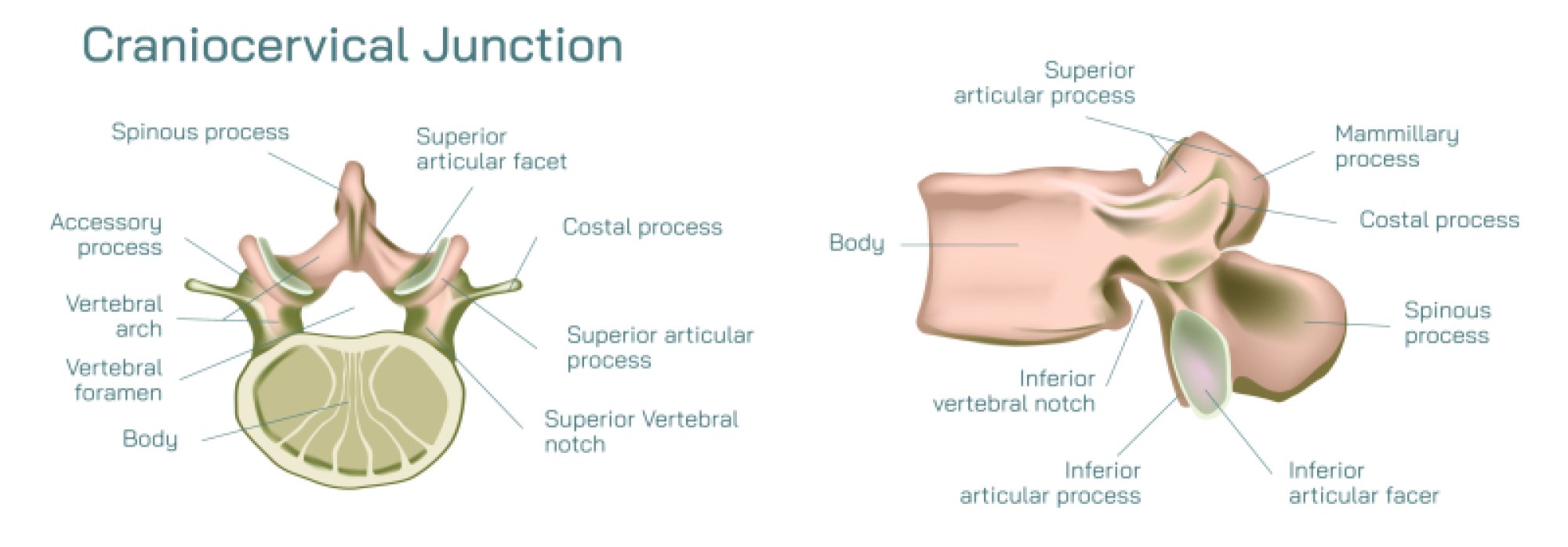
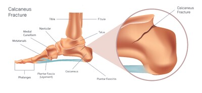
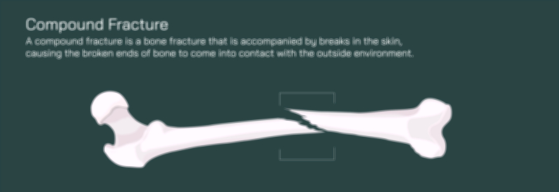
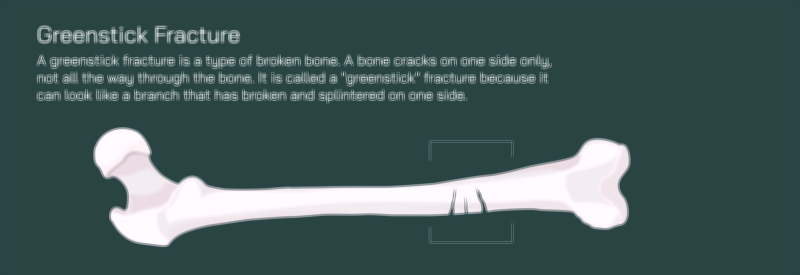
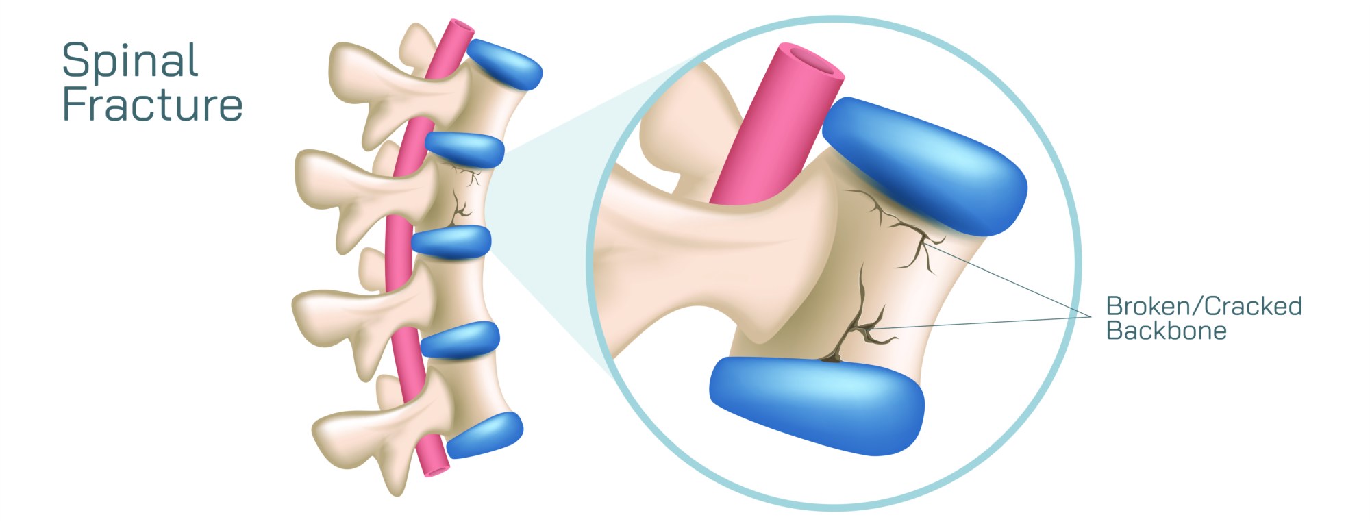
Educational illustrations of spinal cord fractures provide essential visual context for understanding complex anatomy, injury mechanisms, and treatment approaches. These diagrams typically show vertebral alignment, fracture types, ligamentous attachments, and the spatial relationship between bone and spinal cord. Such visual tools are invaluable for medical students, healthcare professionals, and patient education, enhancing comprehension of traumatic spinal injuries, neurological consequences, and therapeutic strategies.