Proximal femur fractures are a major category of skeletal injuries that occur near the upper end of the femur, close to the hip joint. These fractures are commonly referred to as hip fractures and are especially prevalent among elderly individuals due to reduced bone density and increased risk of falls. The classification of proximal femur fractures is primarily based on their anatomical location, as the position of the fracture strongly influences blood supply, healing potential, treatment approach, and overall prognosis. Understanding the different types of proximal femur fractures is essential in anatomy, orthopedics, radiology, and trauma medicine.
The femur is the longest and strongest bone in the human body. Its proximal end consists of the femoral head, femoral neck, greater trochanter, and lesser trochanter, which together form the hip joint and provide attachment sites for powerful muscles. Fractures occurring in this region disrupt both structural stability and mobility, often leading to significant functional impairment. Because the hip joint bears body weight and enables locomotion, injuries in this area require careful clinical assessment and timely intervention.
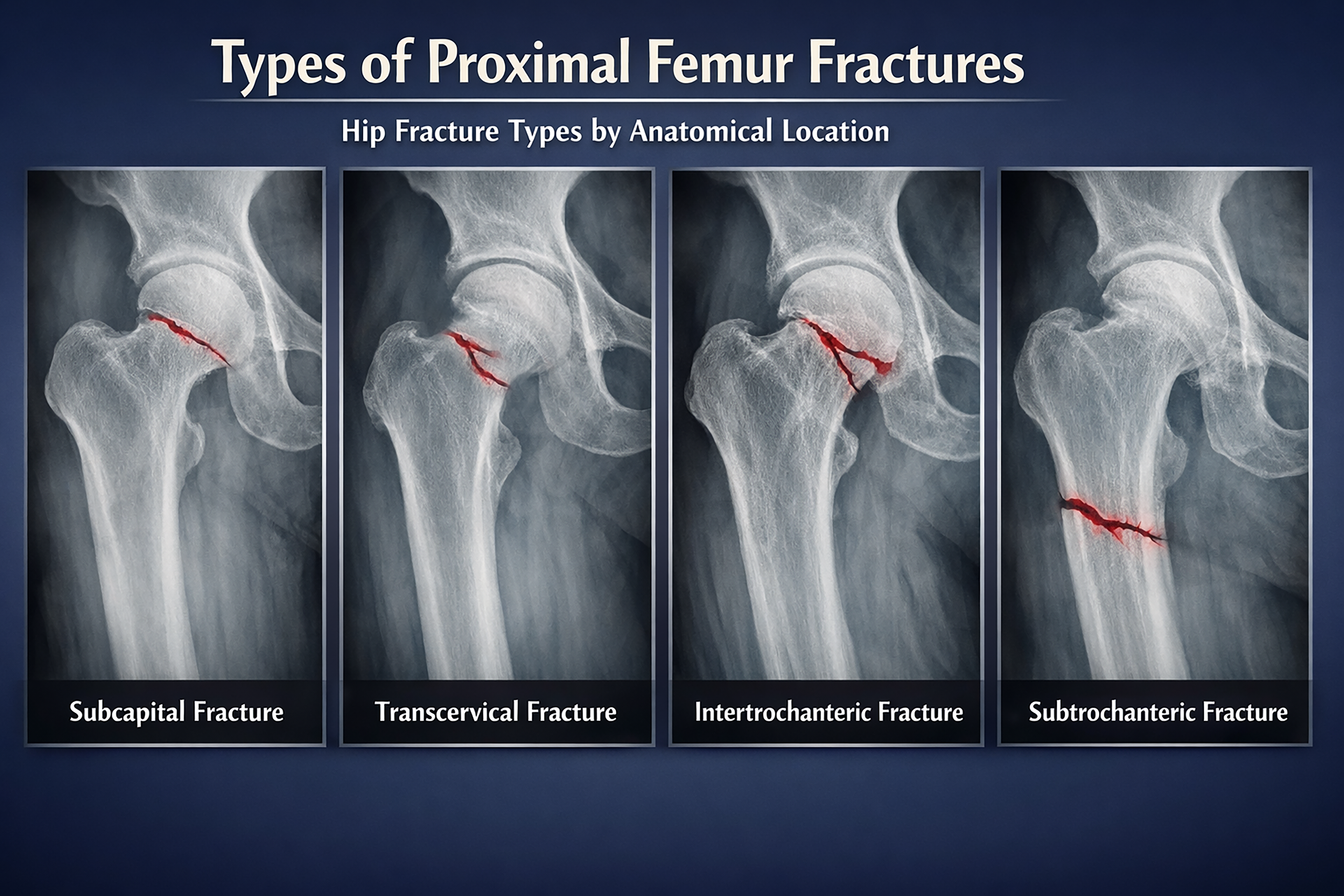
One of the most common types of proximal femur fractures is the subcapital fracture. This fracture occurs just below the head of the femur, at the junction between the femoral head and the neck. Subcapital fractures are intracapsular, meaning they occur within the capsule of the hip joint. This location is clinically significant because it can compromise the blood supply to the femoral head. Reduced blood flow increases the risk of complications such as avascular necrosis and delayed healing. Subcapital fractures are frequently seen in older adults following low-energy trauma such as slipping or falling.
Another important category is the transcervical fracture. This type of fracture occurs through the mid-portion of the femoral neck. Like subcapital fractures, transcervical fractures are also intracapsular and share similar risks related to blood supply disruption. The femoral neck plays a critical role in transmitting forces between the hip joint and the femoral shaft, so fractures in this region can significantly affect mechanical stability. The exact position and displacement of a transcervical fracture influence treatment decisions and long-term outcomes.
Intertrochanteric fractures occur between the greater and lesser trochanters of the femur. These fractures are extracapsular, meaning they lie outside the joint capsule. Because the blood supply to the femoral head is usually preserved, intertrochanteric fractures tend to heal more reliably than intracapsular fractures. However, they often involve significant displacement due to strong muscle attachments in the region. Intertrochanteric fractures are common in elderly individuals and are typically associated with falls or direct trauma to the hip.
Subtrochanteric fractures occur below the lesser trochanter, extending into the upper portion of the femoral shaft. This region experiences high mechanical stress during weight-bearing activities, which can complicate healing. Subtrochanteric fractures may occur due to high-energy trauma in younger individuals or weakened bone structure in older patients. These fractures are often more challenging to manage because of strong muscular forces that cause displacement and delayed union.
The anatomical classification of proximal femur fractures is crucial because each type presents unique clinical challenges. Intracapsular fractures, such as subcapital and transcervical injuries, carry a higher risk of compromised blood flow and long-term complications. Extracapsular fractures, including intertrochanteric and subtrochanteric injuries, generally have better healing potential but may require more complex stabilization due to mechanical forces.
Radiological imaging plays a key role in identifying and classifying proximal femur fractures. X-ray images provide clear visualization of fracture lines, displacement, and anatomical location. Accurate interpretation of these images helps clinicians determine fracture type and plan appropriate management strategies. The diagram showing different proximal femur fracture types serves as a valuable educational tool for understanding these distinctions.
From a clinical perspective, proximal femur fractures are associated with significant morbidity, particularly in older populations. Reduced mobility, prolonged hospitalization, and complications related to immobility can severely affect quality of life. Early diagnosis and proper classification allow for timely treatment, which may include surgical fixation or joint replacement depending on fracture type and patient factors.
Biomechanically, the proximal femur is subjected to complex forces generated by body weight and muscle activity. Fractures disrupt the normal distribution of these forces, leading to pain, instability, and loss of function. Each fracture location alters biomechanics differently, which is why anatomical classification is so important in orthopedic planning.
In medical education, proximal femur fractures are commonly used to teach anatomy, trauma assessment, and fracture management principles. Understanding the differences between subcapital, transcervical, intertrochanteric, and subtrochanteric fractures helps students and healthcare professionals appreciate how anatomy influences clinical decision-making.
Preventive strategies focus on maintaining bone health, particularly in aging populations. Adequate nutrition, physical activity, and fall prevention measures can reduce the risk of proximal femur fractures. Awareness of fracture types also aids in public health education and early intervention efforts.
In conclusion, proximal femur fractures are classified according to their anatomical location into subcapital, transcervical, intertrochanteric, and subtrochanteric types. Each fracture type differs in structural impact, healing potential, and clinical management requirements. Understanding this classification is essential for accurate diagnosis, effective treatment planning, and improved patient outcomes. The diagram illustrating hip fracture types by anatomical location provides a clear and structured overview of these important orthopedic injuries.