Greenstick Fracture of the Femur Bone Showing Partial Crack and Pediatric Bone Injury
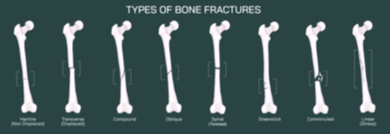
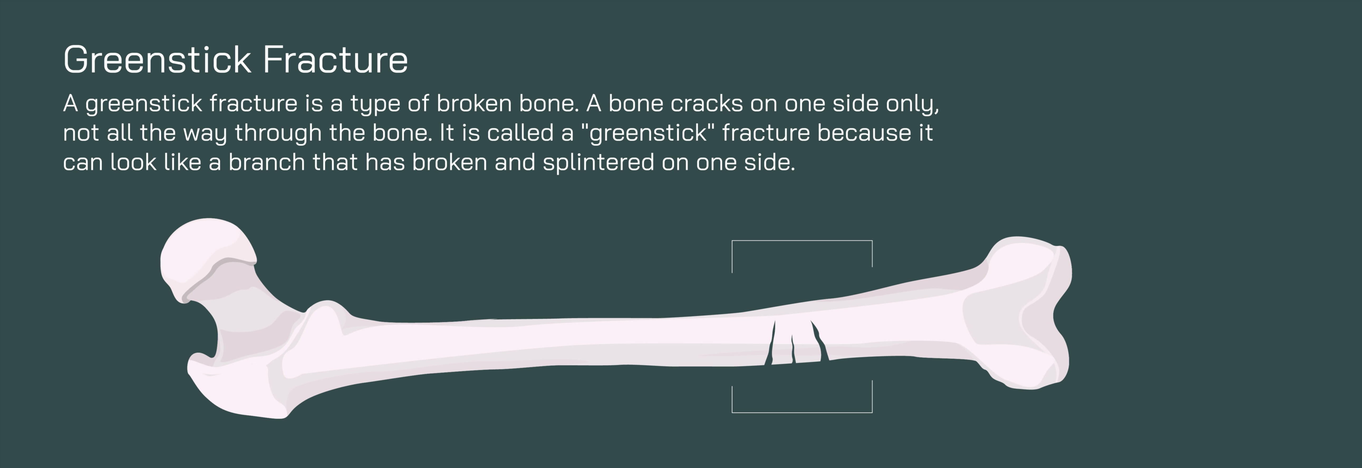
A greenstick fracture of the femur represents a uniquely pediatric pattern of bone injury, one that can occur only in children because of the flexible and developing nature of their skeletal system. Unlike a complete fracture in which a bone breaks fully into two separate pieces, a greenstick fracture involves a partial crack where one side of the bone bends and splinters while the opposite side remains intact. The term “greenstick” comes from the way a fresh, flexible tree branch behaves when bent — it cracks on the outside while the inner side folds without snapping. In a vector illustration, this characteristic pattern is often shown as the femur curving slightly due to the external force, with a visible crack along one cortex of the bone while the remaining cortex remains continuous. This partially broken yet still connected appearance clearly distinguishes a greenstick fracture from other pediatric fractures such as buckle fractures or complete displaced breaks. Because the femur is the largest and strongest bone of the body, even in children, this injury typically results from moderate trauma such as falls while running or jumping, sports collisions, playground accidents, or high-energy impacts like bicycle or skating falls.
Children are especially vulnerable to greenstick fractures because their bones contain a greater proportion of cartilage and collagen fibers compared to adults. Pediatric bone has thick periosteum — the outer protective layer covering bones — and this thick, flexible structure allows bones to bend rather than break cleanly. While this flexibility offers some protection, it can also produce fractures that are easy to underestimate. A child may still be able to bear weight or move the leg, and swelling may be mild, yet a partial fracture can remain hidden beneath the skin. A vector illustration showing the thick periosteal layer surrounding the crack reinforces why these fractures tend to bend instead of separating fully. The illustration makes clear that the outer surface of the bone has broken while the inner surface is held together by soft tissue continuity, a feature that also helps explain why pediatric fractures typically heal rapidly when treated appropriately.
Even though the femur remains structurally connected in a greenstick fracture, the injury is still significant, particularly because the femur supports the full body’s weight during standing and walking. A child with this type of fracture may limp, avoid putting weight on the leg, guard the thigh from movement, or experience pain localized to the crack region. The surrounding soft tissues — including the strong quadriceps and hamstring muscle groups — may tighten reflexively in response to the injury, causing discomfort and difficulty extending or bending the knee. An illustration of the femur surrounded by tightened muscle groups helps convey that although the bone is partially intact, movement is still painful and potentially harmful without stabilization.
Diagnosis often begins with clinical suspicion based on symptoms and mechanism of injury, followed by imaging. X-rays usually reveal the classic pattern: bending of the femoral shaft combined with a clear cortical crack on one side. In some cases, the unbroken side of the bone bows outward, reinforcing the characteristic asymmetry. A pediatric orthopedic illustration may also include comparison images showing how the bone appears pre- and post-reduction, allowing viewers to appreciate how realignment corrects both the crack and the bend.
Because the femur remains partially intact, a greenstick fracture does not self-correct naturally — the bend must be carefully reduced to prevent long-term deformity. If the bone is allowed to heal while bent, the femur may remodel incorrectly, causing leg length discrepancy, altered gait mechanics, or future joint strain. Orthopedic treatment therefore focuses on realigning the bone and holding it in place during healing. For mild cases in which bending is minimal, immobilization with a cast or brace may be sufficient. For more pronounced bending, physicians often perform a gentle manual realignment called closed reduction, sometimes under sedation or anesthesia to keep the child comfortable and ensure accuracy. A vector image that contrasts the misaligned bone before reduction and the straightened bone afterward helps communicate the goal of the procedure — restoring anatomical alignment so that the femur grows normally.
Immobilization typically involves a cast that covers the femur and immobilizes the hip and knee joints to prevent strain on the healing crack. In younger children, a spica cast — which stabilizes the hip and pelvis — may be used. Because pediatric bones heal quickly, the cast period is usually shorter than for adults, and follow-up X-rays monitor progress. As healing progresses, children transition to gradual weight-bearing, gentle range-of-motion exercises, and normal activity as tolerated. A vector diagram illustrating the healing timeline — from initial crack, to early bone bridging, to callus formation, and finally to remodeling — helps show that even a partial fracture undergoes the same biological phases as a complete fracture, just at a faster pace.
The biological advantage of growing bone becomes especially visible in the remodeling phase. Children possess remarkable capacity for reshaping bone over time, often restoring normal structure even after significant bending — provided that the bone is aligned correctly at the start of healing. Osteoblast and osteoclast activity smooths and strengthens the previously cracked cortex, while the periosteum thickens temporarily as part of the healing defense. In vector form, this may be depicted as progressive smoothing and mineral deposition around the crack area until the bone returns to its original cylindrical shape.
Although healing outcomes for greenstick femur fractures are excellent when treated promptly, delayed or improper management can lead to complications. Persistent angulation, rotational deformity, leg length differences, muscle imbalance, and recurring pain during growth spurts are possible risks. Illustrations that juxtapose untreated and properly treated bone make the importance of orthopedic follow-up intuitive and memorable to families.
The psychological component of pediatric fractures is also important. Children often recover physically faster than they do emotionally; fear of re-injury or hesitancy to resume activities is common. A vector educational image showing a timeline of safe return to play — rest, recovery, early walking, running, and eventual full sports — helps reassure families that temporary restriction leads to long-term protection and healthy musculoskeletal development.
In its fullest educational purpose, a vector illustration of a greenstick fracture of the femur showing partial crack and pediatric bone injury weaves multiple ideas into one visual story: the unique flexibility of children’s bones, the bending-and-splintering fracture pattern, the orthopedic correction needed to restore alignment, and the rapid healing capacity of growing skeletons. It demonstrates that pediatric fractures are not simply “smaller versions” of adult fractures; they follow their own biomechanical rules and require tailored treatment. By showcasing both the partial crack and the healing pathway, the illustration becomes an invaluable tool for parents, clinicians, medical students, physical therapists, and anyone seeking to understand how childhood bone injuries behave and how expert orthopedic care ensures healthy growth and lifelong mobility.