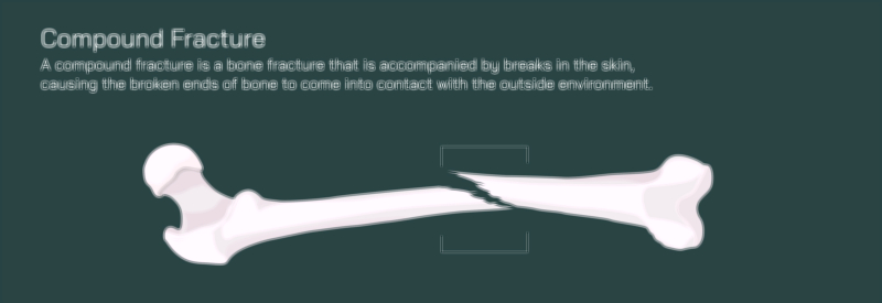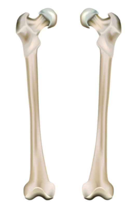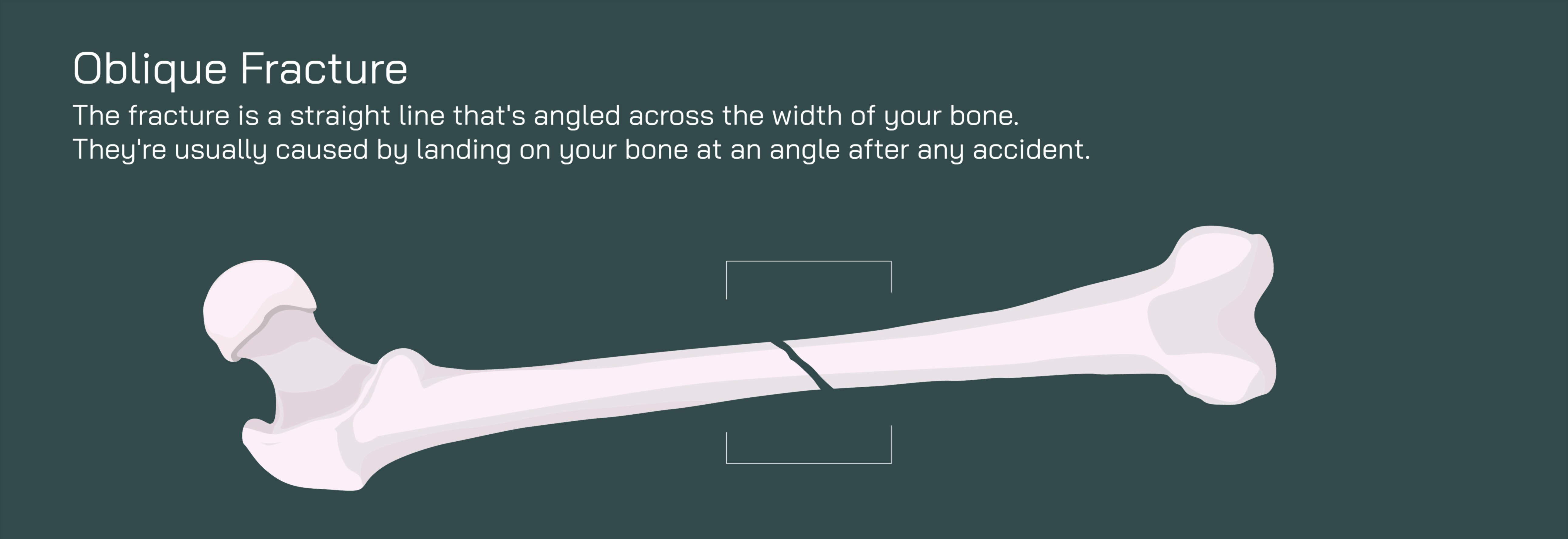
Oblique Fracture of the Femur Bone Showing Diagonal Crack and Orthopedic Healing Alignment
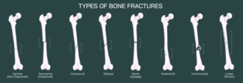
An oblique fracture of the femur is a distinctive type of bone break in which the fracture line runs diagonally across the shaft, forming an angled separation rather than a straight horizontal or vertical crack. This angled pattern is produced when the femur is exposed to a combination of compression and rotational forces — for example, when the leg is twisted during impact, caught in a fall, or subjected to a sudden change in direction under heavy load. A vector illustration of this injury typically depicts the femoral cortex split along a slanted plane, forming two main fragments with sharply angled edges. While the femur is the strongest long bone in the body, capable of supporting tremendous weight and force distribution, an oblique fracture disrupts its mechanical stability in a way that allows the bone ends to slide across one another. This sliding tendency is a defining feature of oblique fractures and one of the main reasons orthopedic stabilization is required.
Externally, an oblique fracture may present with dramatic symptoms: intense pain, rapid swelling, difficulty or inability to bear weight, shortening or visible deformity of the leg, and involuntary muscle spasms that worsen displacement. The strong thigh muscles — particularly the quadriceps, hamstrings, and adductors — pull unevenly on the diagonal fragments, causing them to shift out of alignment. A vector representation showing muscle tension and fragment shifting helps explain why this fracture cannot remain stable without medical intervention. Even if the skin remains intact, an oblique fracture is mechanically unstable and can quickly progress from a clean angled break into a displaced or overlapping fracture if weight-bearing continues.
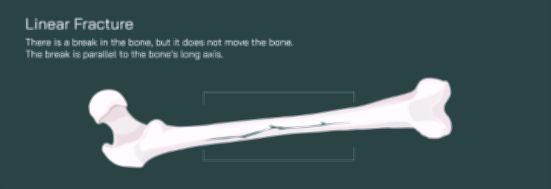
Radiographic imaging confirms the diagnosis by revealing the classic angled fracture line. The fracture may pass through the mid-shaft of the femur or involve areas closer to the hip or knee, and its length and angle vary depending on the mechanism of injury. Illustrations often label the fracture plane while emphasizing how easily the slanted edges can slip over one another when unsupported. This visual cue is crucial for understanding why a fracture with just two fragments can still be more unstable than a linear fracture with a single crack.
Because of this inherent instability, orthopedic treatment for an oblique femur fracture is designed to restore proper alignment and prevent fragment slippage throughout healing. The process begins with reduction, the realignment of the broken bone ends into their normal anatomical position. Reduction may be achieved through manual repositioning, controlled traction, or surgical manipulation depending on the severity of displacement. A vector illustration that shows the before-and-after alignment of the angled fragments makes the purpose of reduction immediately clear — restoring the continuous long-axis shape of the femur.
Once alignment is established, stabilization ensures that the fragments remain locked in position while the body repairs the break. The preferred method for most oblique femur fractures is intramedullary nailing, in which a metal rod is inserted into the central marrow canal and secured with locking screws above and below the fracture site. Because the rod sits inside the bone, it resists rotational and sliding forces far better than a cast alone. A vector diagram that shows the nail crossing the fracture line helps viewers visualize the internal scaffolding responsible for maintaining stability.
In fractures that require additional support — such as when the fracture angle is steep or the bone quality is low — surgeons may add plates and screws along the outer bone surface to anchor the fragments. For cases involving severe trauma or swelling, external fixation may be used temporarily, consisting of metal pins connected to an external stabilizing frame. A labeled illustration comparing internal and external fixation methods is especially useful for educational purposes, demonstrating how each mechanism counters rotational shear and sliding forces produced by the oblique fracture plane.
The biological healing sequence follows the familiar four phases of bone recovery:
Inflammatory Phase (first days): Blood pools around the fracture to form a hematoma that triggers immune and repair cell activation.
Soft Callus Phase (weeks 2–6): Collagen and cartilage begin bridging the angled fracture gap, stabilizing the fragments internally.
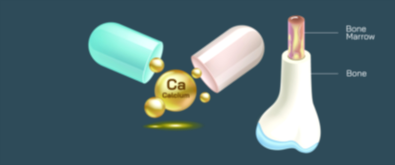
Hard Callus Phase (weeks 6–12): Calcium and phosphorus mineralization convert the soft callus into hardened bony tissue, strengthening the repair.
Remodeling Phase (months–years): The bone reshapes and reinforces itself, gradually restoring strength and smoothing excess callus to match its original form.
Vector illustrations that depict this sequence — from the slanted fracture line, to early callus formation, to final bone remodeling — help clarify both the timescale and the internal changes occurring even while the external leg appears healed.
Rehabilitation begins when imaging confirms early callus stability. Weight-bearing progresses gradually to stimulate bone strengthening without jeopardizing alignment. Physical therapy restores hip and knee mobility, rebuilds quadriceps and hamstring strength, and corrects gait abnormalities caused by pain or immobilization. Since improper loading can cause the diagonal fracture to slip before complete healing, vector illustrations showing the transition from crutches to full ambulation emphasize the importance of controlled progression.
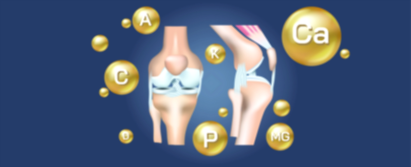
Nutrition and systemic health significantly influence healing outcomes. Adequate calcium, vitamin D, magnesium, and protein support mineralization and collagen formation, while smoking, alcohol, and poor diet delay healing. For individuals with low bone density, medications that enhance bone formation may be recommended to reduce the risk of delayed union or refracture.
Although oblique fractures of the femur typically heal well with proper orthopedic care, complications can arise if alignment is not restored or maintained. Malunion — healing in an incorrect position — can result in leg length differences, altered gait mechanics, chronic pain, and increased joint wear over time. A vector comparison of a properly aligned femur versus a malaligned healed femur highlights why follow-up imaging, adherence to mobility guidelines, and physical therapy are critical.
In its full educational purpose, a vector illustration of an oblique fracture of the femur showing a diagonal crack and orthopedic healing alignment provides more than a view of the broken bone — it tells the entire story of injury, treatment, and recovery. It visually explains why angled fractures are unstable, why internal fixation is typically required, how biological healing progresses, and how rehabilitation restores mobility. By making the invisible process of alignment-dependent healing visible, the illustration becomes an effective tool for medical learners, orthopedic professionals, physical therapists, patients, and families — reinforcing that femur fractures demand respect, precision, and patience to return safely to full strength and active movement.