Spiral Fracture Showing Twisting Break Pattern in a Long Bone and Orthopedic Healing Alignment
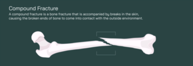
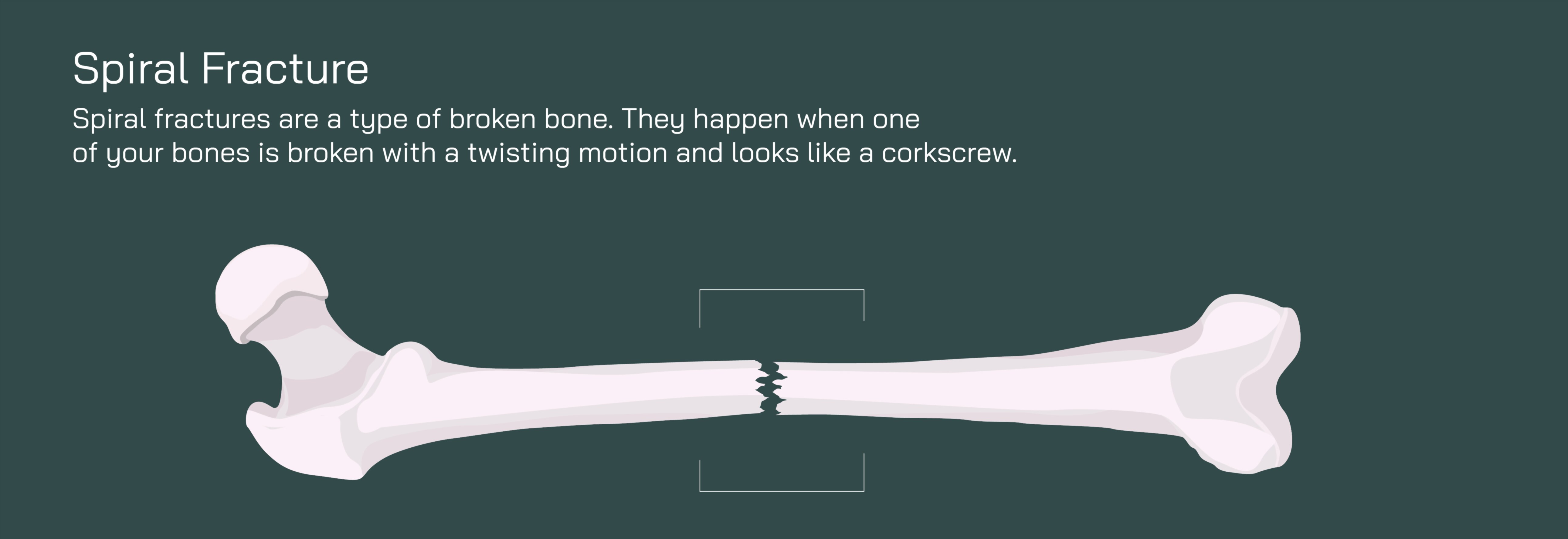
A spiral fracture is a dramatic and mechanically complex injury in which a bone breaks along a twisting or corkscrew-like path, produced by rotational force rather than direct impact alone. In this pattern, the fracture line winds diagonally around the shaft of a long bone — such as the femur, tibia, humerus, or fibula — instead of running straight across or lengthwise. A vector illustration of a spiral fracture typically shows the bone intact in overall structure but split in a helical pattern, resembling a stripe spiraling around the bone. This visual immediately conveys that the injury originates from torsion, the same mechanical principle that occurs when a solid object is twisted until it cracks along a diagonal–rotational plane. In everyday life, spiral fractures are often the result of forceful twisting during sports, skiing accidents, falls in which one limb gets trapped while the body spins, industrial injuries, or accidents involving sudden rotational trauma.
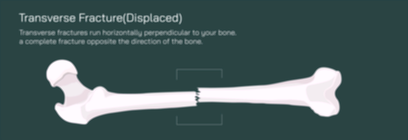
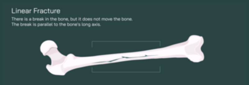
One of the defining features of a spiral fracture is its instability. Because the break follows a slanted and rotating path, the fractured ends tend to slide and rotate relative to each other. Unlike a linear fracture — which typically preserves alignment — a spiral fracture disrupts the bone’s natural structural geometry. Powerful surrounding muscles may involuntarily pull the broken ends into different rotational positions, causing pain, deformity, and limb misalignment. A vector illustration that highlights both the helical break and the directional pull of muscle forces helps learners understand why spiral fractures require more than immobilization; they demand precise orthopedic correction to control rotation.
Externally, symptoms often include immediate sharp pain, swelling, bruising, inability to bear weight, and limb deformity — sometimes appearing as an abnormal inward or outward rotation of the leg or arm. Because rotational displacement is less visually obvious than bending displacement, imaging such as X-rays — and in complicated cases, CT scans — play a critical role. Radiographs reveal the tell-tale corkscrew-shaped fracture line, confirming the diagnosis and guiding treatment decisions. Vector images that overlay the twist direction and fracture shape on the bone offer a clear explanation of the injury mechanism.
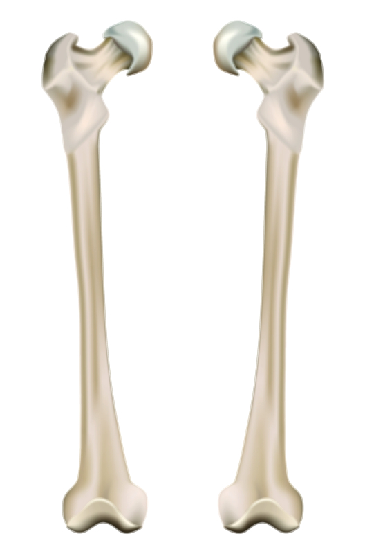
Orthopedic healing alignment for a spiral fracture centers on three goals: correcting rotation, restoring anatomical length, and stabilizing the bone to prevent re-twisting during healing. Treatment begins with reduction, a manual or surgical process in which the orthopedic team realigns the broken bone ends into their exact original orientation. Because misalignment at the rotational level can cause long-term disability, reduction must be highly precise — even a few degrees of rotational error can produce gait abnormalities, joint wear, or chronic pain. Illustrations showing “before and after” rotational correction make the purpose of reduction visually clear.
Once proper alignment is achieved, stabilization prevents the spiral edges from slipping. This is most commonly accomplished through internal fixation, especially in weight-bearing long bones like the femur or tibia. The gold-standard method is intramedullary nailing, in which a long metal rod is inserted through the marrow canal and secured with locking screws at both ends of the fracture. The rod resists rotational torque and prevents the twisted segments from shifting. A vector diagram showing the rod running through the break helps learners visualize this internal support mechanism.
In some cases — particularly when the spiral fracture includes multiple secondary fragments or involves a joint — plates and screws may be applied along the outer cortex of the bone to secure the helical pattern and eliminate rotational play. For high-energy trauma with extensive soft-tissue damage, external fixation may temporarily stabilize the bone until internal fixation becomes safe. A side-by-side vector comparison of internal versus external fixation methods helps clarify how different hardware designs counter twisting forces.
The body then performs the biological repair sequence, and healing can be illustrated in four stages:
Inflammatory Phase (first days): A hematoma forms around the spiral break, bringing immune cells to clear debris and prepare the site for repair.
Soft Callus Phase (weeks 2–6): Collagen and fibrocartilage begin bridging the helical gap. Although the bone is still fragile, the fragments start to unify.
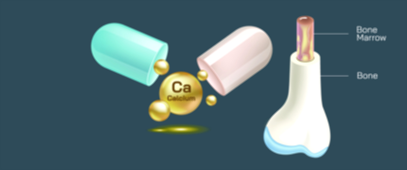
Hard Callus Phase (weeks 6–12): Mineralization — primarily with calcium and phosphorus — strengthens the soft callus into solid new bone. The spiral line becomes less prominent on imaging.
Remodeling Phase (months–years): Bone gradually reshapes, restoring its cylindrical structure and internal alignment to match mechanical stress patterns. The fracture becomes nearly indistinguishable from surrounding bone.
A visual timeline showing callus formation wrapping around the bone in a spiral pathway illustrates how the healing pattern mirrors the original injury route.
Rehabilitation begins when early healing is confirmed radiographically. Gradual return to weight-bearing and controlled mobility helps stimulate bone strengthening without jeopardizing alignment. Physical therapy rebuilds surrounding muscle strength, restores joint range of motion, and retrains gait mechanics to prevent long-term abnormalities. Vector illustrations that track the progression from crutches to full weight-bearing support understanding of safe healing.
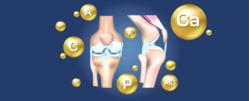
Nutrition and metabolic support strengthen the orthopedic healing process. Calcium, vitamin D, magnesium, protein, and balanced hydration play critical roles in callus formation and mineralization. For individuals with low bone density or recurrent fractures, medications that support bone metabolism may be recommended. A graphic overlay of nutrient icons feeding the healing callus helps tie biological chemistry to orthopedic recovery.
If rotational alignment is not corrected or stabilized sufficiently, complications can arise — including malunion (twisted healing), chronic pain, joint strain, gait irregularities, delayed union, or nonunion. A visual comparison of correct versus incorrect alignment after healing drives home why follow-up imaging, adherence to movement restrictions, and structured rehabilitation are essential.
Ultimately, a spiral fracture vector illustration showing the twisting break pattern and orthopedic healing alignment does far more than display a broken bone. It tells a complete story — how rotational force produces a helical fracture, why this pattern is inherently unstable, how orthopedic treatment restores precise alignment, how internal hardware provides mechanical resistance to twisting, and how the body rebuilds bone through staged biological healing. By visually pairing the injury mechanism with the recovery pathway, the illustration becomes a clear and powerful tool for clinicians, medical students, physical therapists, patients, and families — reinforcing that successful healing depends on precision, protection, and time for the body’s natural repair system to rebuild strength and function.