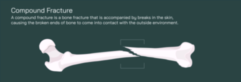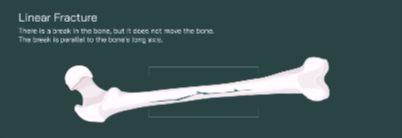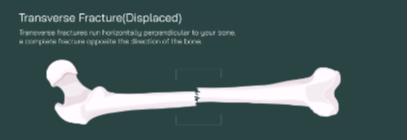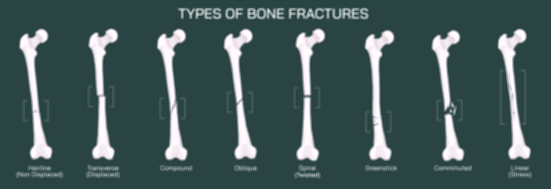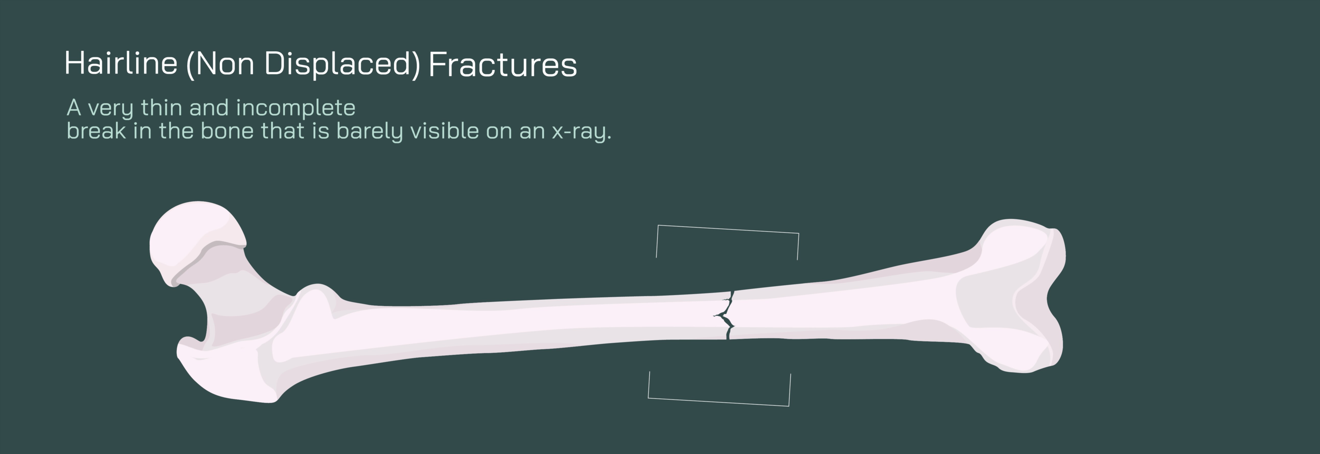
Hairline Non-Displaced Fracture of the Femur Bone Showing Crack and Healing Process
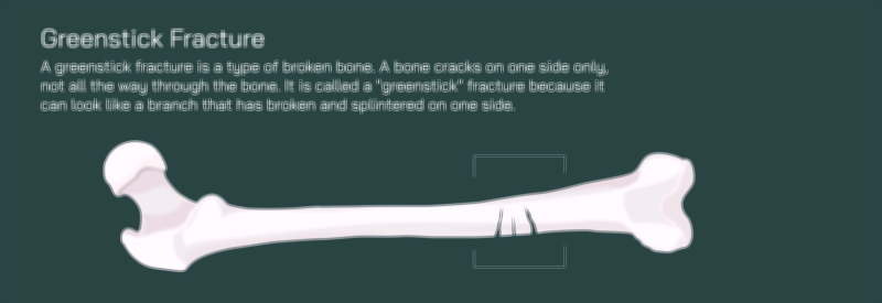
A hairline non-displaced fracture of the femur is one of the most subtle yet medically significant injuries that can occur within the skeletal system. Unlike dramatic fractures where a bone breaks completely or shifts out of alignment, a hairline fracture — sometimes called a stress fracture or micro-fracture — involves a very thin crack in the bone without any separation or dislocation of the fractured segments. In a vector illustration, this type of injury is depicted by showing the femur bone intact in its normal anatomical shape, but with a narrow, fine crack line running across or along the bone surface. Because the bone fragments remain perfectly aligned, the femur retains its structure, and there is no visible deformity from the outside. Yet beneath the skin, the injury represents a failure of the bone’s internal micro-architecture caused by repetitive stress, overuse, reduced bone density, or sudden spikes in physical loading. This form of fracture requires careful diagnosis and thoughtful management to prevent the crack from widening into a complete displacement.
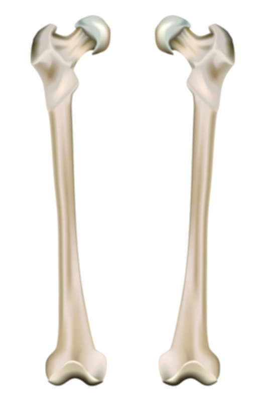
The femur — the strongest and longest bone in the body — supports the entire weight of the torso and transfers forces between the hip and the knee. Even though it is built for heavy load-bearing, repeated high-impact activities such as long-distance running, jumping sports, sudden increases in training intensity, or poor biomechanics can overwhelm the bone’s ability to remodel itself quickly enough. In individuals with weakened bone density due to osteoporosis, nutritional deficiencies, hormonal imbalances, or insufficient calcium intake, even everyday activities can generate enough stress to produce a crack. A vector illustration of the femur showing internal trabecular tissue alongside the crack helps communicate that the injury begins at the microscopic level long before symptoms make it detectable.
Because non-displaced hairline fractures do not deform the bone, diagnosis can be difficult with ordinary physical examination. There may be no visible bruising or swelling; instead, patients often report deep, pinpoint pain that worsens with pressure or weight-bearing and improves with rest. Early X-rays sometimes miss hairline cracks because mineral loss and callus formation take time to appear on imaging, so MRIs or bone scans are often needed for confirmation. A visual illustration that overlays the crack on a realistic femur silhouette can highlight why clinical suspicion matters even when imaging results are initially inconclusive.
Healing of a hairline femur fracture is a multi-stage biological repair process that depends heavily on immobilization, blood supply, and nutrient availability. A vector visual showing the timeline of healing captures the progression clearly:
• Inflammatory Phase (first days) — The body reacts immediately after injury by sending blood and immune mediators to the fracture site. Although the crack is small, a hematoma forms around it, which contains the first building blocks needed for repair. In an illustration, this may appear as a mild discoloration or swelling around the crack.
• Soft Callus Phase (weeks 2–6) — New connective tissue and fibrous cartilage bridge the crack, stabilizing the fracture internally. The bone has not yet regained its full strength, but the edges are prevented from moving apart. A vector diagram often shows this callus as a cloudy band linking both sides of the crack.
• Hard Callus Phase (weeks 6–12) — Calcium and phosphorus begin to mineralize the soft callus, gradually converting it into hardened bone tissue. In an illustration, this appears as a thickened, whiter, denser segment surrounding the original crack, indicating reinforcement.
• Remodeling Phase (months to years) — Osteoclasts and osteoblasts reshape the newly formed bone, smoothing and reorganizing it to match the original femur structure. Over time, the repaired region becomes nearly indistinguishable from the surrounding bone. In a vector depiction, the crack line disappears as uniform bone texture returns.
A key advantage of non-displaced hairline fractures is that, because the bone alignment remains intact, the risk of surgical intervention is lower. Most cases heal with rest, restricted weight-bearing, immobilization, and gradual return to activity. Crutches or reduced activity are often necessary to protect the bone while callus formation progresses. If the individual resumes high-impact activities too soon, the micro-crack can widen and become a full displaced fracture requiring surgical fixation with rods, screws, or plates. This is why educational diagrams commonly include caution arrows showing the danger of premature stress on the healing bone.
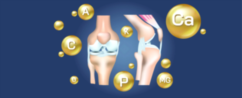
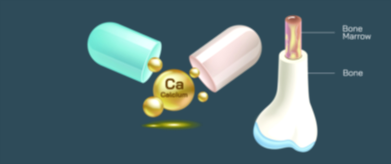
Healing also depends greatly on bone nutrition and metabolism. Adequate calcium intake, vitamin D for absorption, and magnesium for stabilization help the body deposit new tissue effectively during callus formation. Protein supports collagen synthesis, while overall hydration and blood circulation assist in delivering nutrients to the healing site. In older adults, hormonal support and bone-density monitoring may also be necessary to prevent recurrence.
A vector illustration showing the healing stages of a hairline fracture of the femur not only demonstrates the crack itself but also highlights what the body does to restore strength and continuity. When viewers see the femur before injury, during the fracture, during callus development, and after remodeling, the invisible process becomes easy to understand. For clinicians, physical therapists, sports trainers, and patients, such an illustration becomes an educational roadmap that explains why rest, nutrition, and gradual rehabilitation are essential — even when the fracture is tiny and painless at rest.
Ultimately, thinking of a hairline non-displaced femur fracture visually reinforces a crucial truth: bone injuries do not need to be dramatic to be serious. Even a nearly invisible crack represents a break in the structural integrity of the body’s strongest bone, and only patience, biological healing, and proper recovery can restore full strength and mobility.