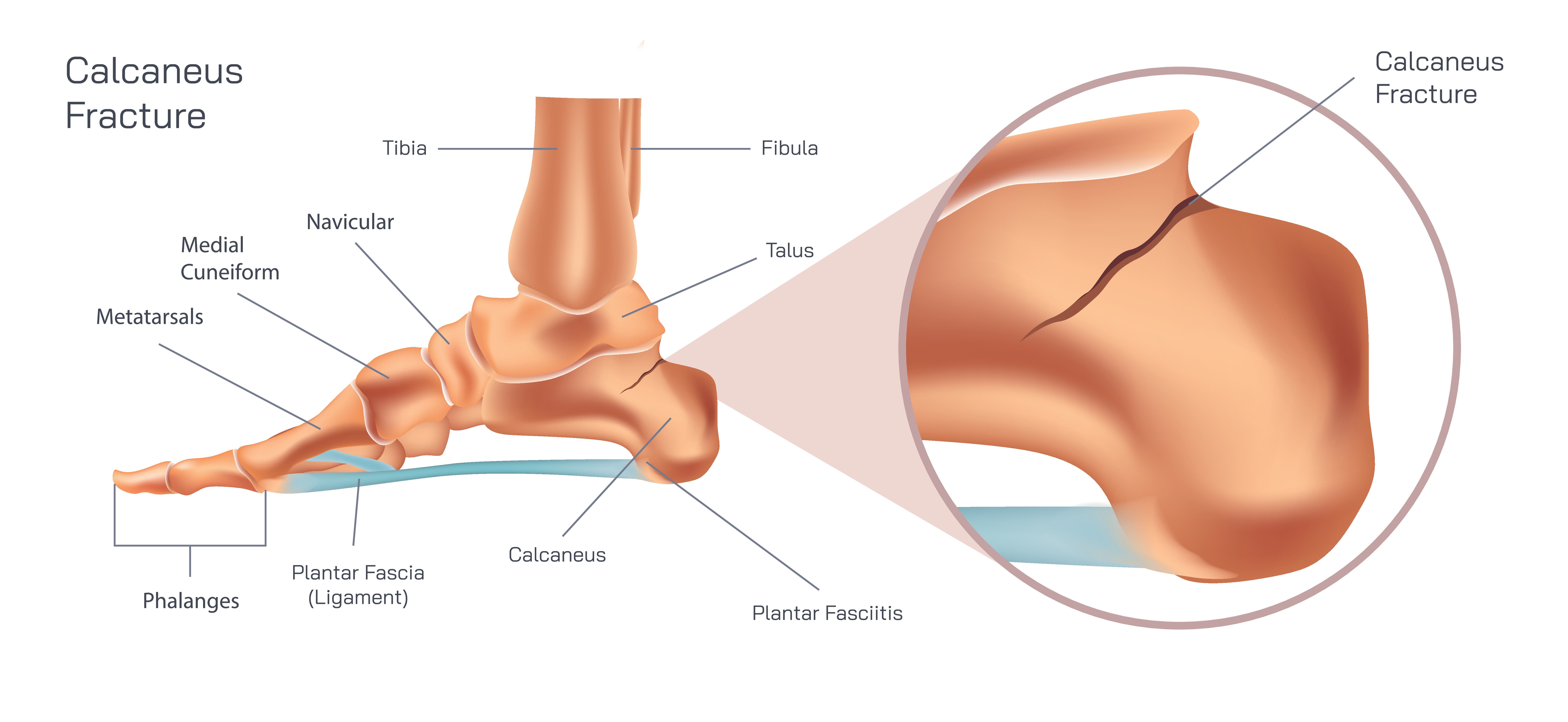
A calcaneus fracture, commonly referred to as a broken heel bone, is one of the most serious injuries of the foot because the calcaneus plays a crucial role in weight bearing, shock absorption, and locomotion. The heel is the first part of the foot to contact the ground during walking, and it absorbs a large portion of the body’s total impact during movement. When the calcaneus fractures, the structure of the heel becomes disrupted, affecting alignment of the subtalar joint and the biomechanics of the entire lower limb. Even though the heel is a dense and powerful bone, the force required to break it is usually considerable, often resulting from high-energy trauma such as falls from height, road accidents, sports injuries, or severe twisting impact. The result can range from a clean break to a comminuted fracture in which the bone shatters into multiple pieces, creating lifelong implications if not managed properly.
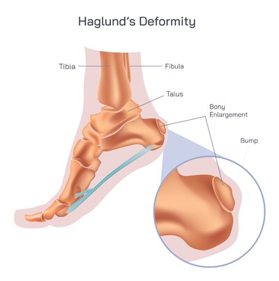
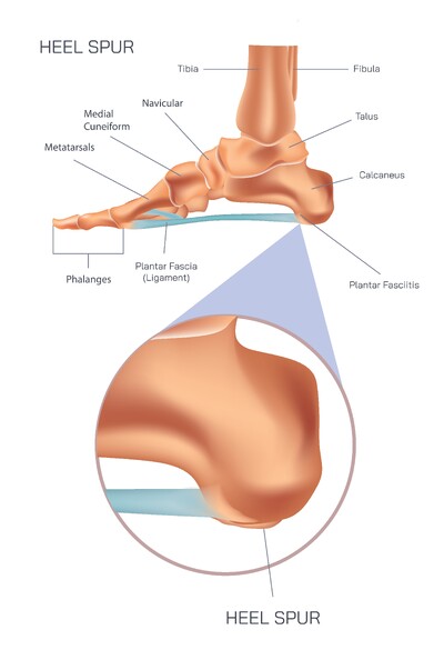
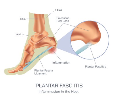
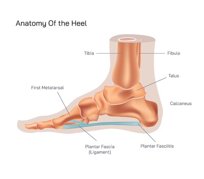
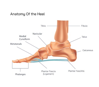
The calcaneus is located at the back of the foot and forms the foundation of the hindfoot. It articulates superiorly with the talus at the subtalar joint and anteriorly with the cuboid bone. This joint configuration is responsible for side-to-side motion of the foot, enabling adaptability during walking. When a calcaneus fracture occurs, the smooth joint surface can become damaged or displaced, compromising joint mobility and increasing the risk of arthritis if not treated correctly. The fracture often causes the heel to widen, shorten, or change shape due to compression of the bone, which then alters body posture and gait. For this reason, calcaneus fractures are not only painful but also biomechanically disruptive.
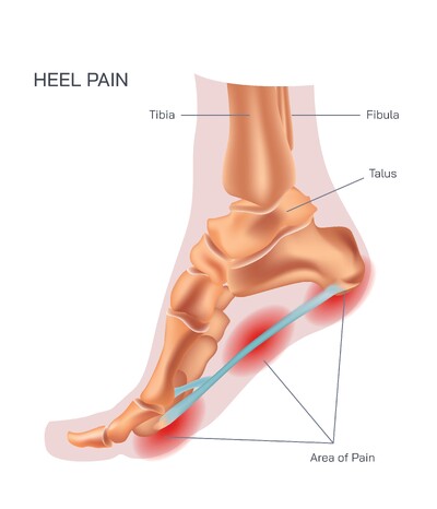
Symptoms of a calcaneus fracture typically appear immediately after the injury and include intense heel pain, rapid swelling, bruising of the foot and ankle, inability to bear weight, and noticeable deformity in severe cases. Because the heel controls shock absorption, any attempt to stand or walk amplifies pain dramatically. Patients may also report numbness or tingling due to nerve compression or tight compartment pressure. Given the strong impact required to fracture the calcaneus, associated injuries such as lumbar spine trauma and tibial fractures are not uncommon, and this is why comprehensive assessment is essential.
Diagnosis relies on physical examination followed by imaging. X-rays can reveal the type and extent of fracture, while computed tomography (CT scanning) provides a detailed three-dimensional view of bone fragments and joint involvement. When soft-tissue injury is suspected, magnetic resonance imaging (MRI) may also be used. Physicians assess whether the fracture is displaced, non-displaced, open, intra-articular, or extra-articular, because classification determines treatment strategy and prognosis.
Treatment varies widely depending on severity. Non-displaced fractures may be managed conservatively through immobilization, rest, elevation, ice, casting, splinting, and long-term non-weight bearing. In these cases, healing can progress without altering bone shape, although recovery remains slow and gradual. Displaced or comminuted fractures often require surgical intervention to restore anatomical structure. Surgical procedures may involve open reduction and internal fixation (ORIF) using plates and screws to realign the bone and joint surface. Some fractures with severe joint destruction may require subtalar fusion to prevent disabling chronic pain. Surgeons must also consider swelling, skin damage, and circulation around the heel before attempting procedures because the surrounding soft-tissue environment is vulnerable to complications.
Recovery from calcaneus fractures is lengthy because the heel is a weight-bearing bone and cannot be stressed prematurely without risking reinjury or malunion. Patients typically remain non-weight bearing for several weeks or months before gradually progressing to partial and then full weight bearing under medical supervision. Physical therapy becomes critical during this stage to restore range of motion, reduce stiffness, rebuild muscle strength, and regain proper gait. The subtalar joint is particularly susceptible to long-term stiffness, so early and consistent rehabilitation helps prevent future limitations.
Pain management, specialized footwear, cushioning inserts, and progressive loading strategies support everyday function as healing progresses. Even with proper treatment, some individuals continue to experience chronic heel pain, decreased ankle mobility, altered gait, or early arthritis. However, committed rehabilitation and supportive care greatly improve long-term outcomes. For athletes, labor workers, and active individuals, structured physical conditioning and gradual return to high-impact activities reduce reinjury risk and protect the heel from overloading.
A calcaneus fracture is far more than a simple broken bone; it is an injury that affects the mechanics of the entire lower limb. Understanding the anatomy of the heel, the complexity of the joint surface, and the importance of precise realignment helps highlight the significance of timely diagnosis and targeted treatment. Patients who prioritize rehabilitation and long-term support of heel biomechanics often regain mobility and resume a fulfilling lifestyle. With medical attention, structured recovery, and thoughtful preventive habits, individuals can protect the heel from further trauma and maintain healthy movement for years ahead.