Spinal Cord Craniocervical Junction – Anatomy, Function, and Clinical Significance
The craniocervical junction (CCJ) represents a critical anatomical region where the brainstem transitions into the spinal cord, connecting the skull base to the upper cervical vertebrae, primarily the atlas (C1) and axis (C2). This junction is a complex interface that supports both structural stability and neurological function, integrating motor, sensory, and autonomic pathways essential for life-sustaining processes. Its unique anatomy allows the head to rotate, flex, and extend while safeguarding delicate neural structures.
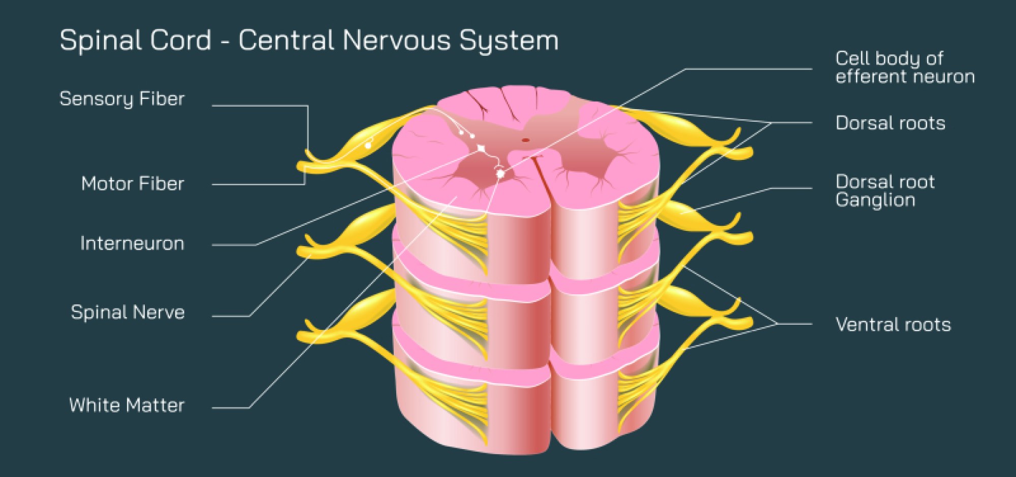
The spinal cord at the craniocervical junction contains bundles of ascending and descending nerve fibers that transmit sensory information to the brain and motor commands to the body. This includes critical pathways controlling respiration, cardiovascular regulation, and limb movement. The CCJ is surrounded by ligaments, cerebrospinal fluid, and vertebrae that protect the cord while allowing flexibility and movement.
Clinical significance of the craniocervical junction is profound. Trauma, congenital malformations, tumors, or degenerative diseases in this area can result in neurological deficits, including weakness, sensory loss, or even respiratory compromise. Understanding the detailed anatomy of the CCJ is essential for neurosurgeons, radiologists, and neurologists when planning surgical interventions, imaging interpretation, or assessing spinal stability.
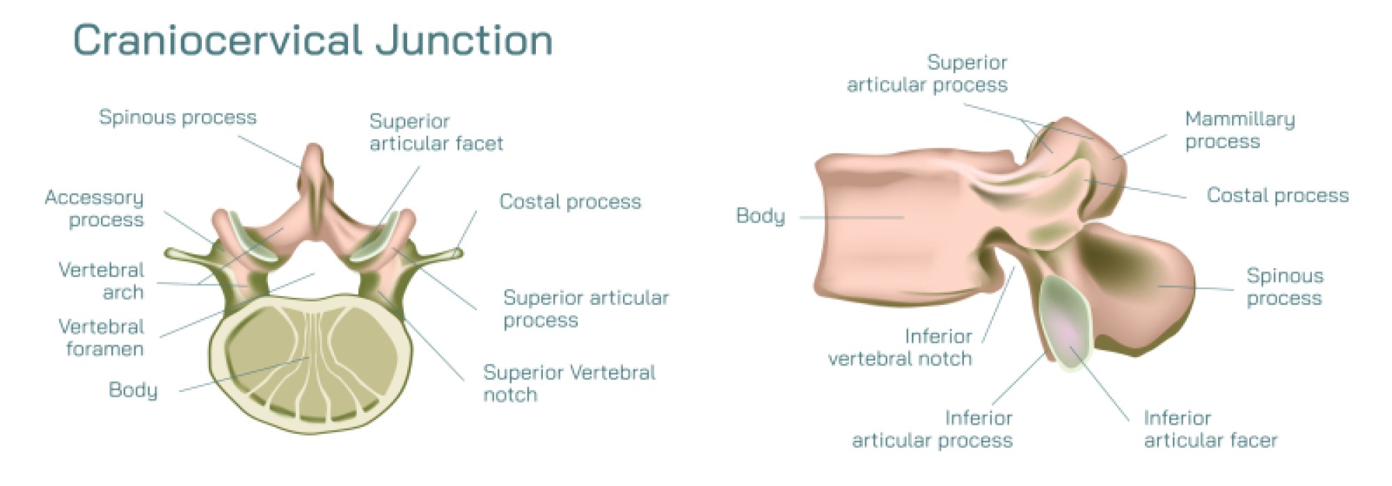
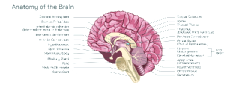
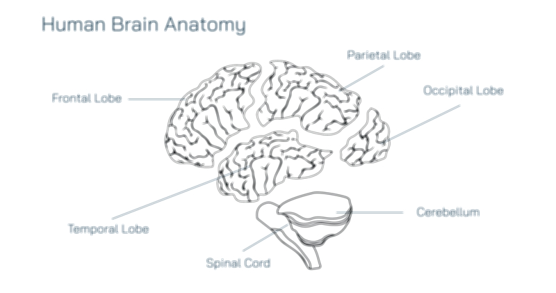
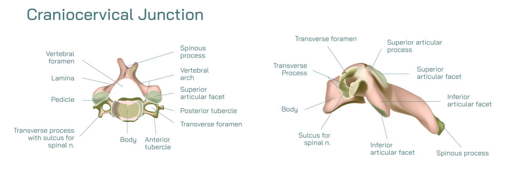
Educational illustrations and 3D visualizations of the spinal cord craniocervical junction are invaluable for learning and clinical training. Diagrams typically show the brainstem, medulla oblongata, upper cervical spinal cord, atlas and axis vertebrae, and emerging spinal nerves. Highlighting the alignment and relationships between bony structures, ligaments, and neural pathways helps students and professionals appreciate both anatomy and function.
Vector and 3D illustrations also allow visualization of nerve pathways, cerebrospinal fluid flow, and ligament support, providing insight into how trauma or compression can impact neurological function. These resources are essential in medical education, patient counseling, and research, enabling a clear understanding of structural and functional interconnections.
In conclusion, the spinal cord craniocervical junction is a pivotal anatomical region bridging the brainstem and cervical spine. Its structural complexity and neurological importance make it a focus of medical education, clinical evaluation, and surgical planning. High-quality illustrations and 3D models enhance comprehension of this junction, making it accessible for learners, healthcare professionals, and researchers, while emphasizing its role in maintaining stability, mobility, and vital neural functions.