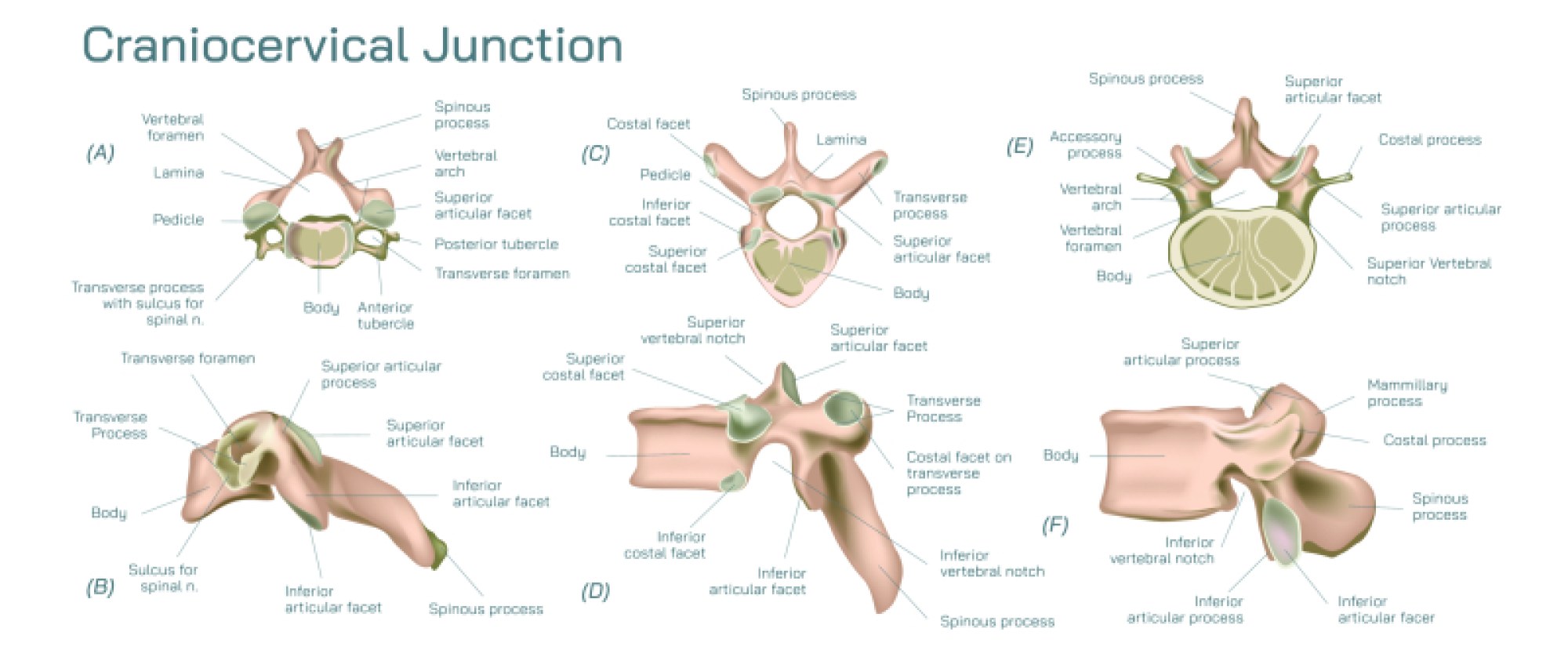
Educational Overview of the Craniocervical Junction
The craniocervical junction represents a complex anatomical region where the base of the skull meets the upper cervical spine. This junction includes critical structures such as the occiput, atlas (C1), and axis (C2) vertebrae, which together provide both support and mobility to the head. The foramen magnum, a prominent opening in the occipital bone, allows passage of the brainstem and upper spinal cord. The unique articulation between atlas and axis facilitates pivotal head movements, including rotation, flexion, and extension, making this region crucial for functional mobility and neuromuscular coordination.
Several ligaments stabilize the craniocervical junction. The transverse ligament of the atlas holds the dens (odontoid process) against the anterior arch, preventing excessive movement that could compromise the spinal cord. The alar ligaments connect the dens to the occipital condyles, limiting rotation and side-bending. The apical ligament extends from the tip of the dens to the anterior margin of the foramen magnum, providing additional vertical stabilization. Together, these ligaments maintain alignment, protect neural structures, and distribute mechanical stress during head movements.
From a clinical perspective, understanding this junction is critical for diagnosing and managing trauma, degenerative changes, congenital anomalies, and inflammatory conditions. Injuries such as atlantoaxial instability, odontoid fractures, or ligamentous disruption can have severe neurological consequences. Radiological imaging, including CT and MRI, often relies on detailed anatomical knowledge of the craniocervical junction to evaluate pathology and guide surgical interventions.
Educational illustrations of this area typically depict bones, ligaments, joints, and neurovascular pathways in clear, labeled diagrams. These visual tools are indispensable for medical students, healthcare professionals, and educators seeking to convey complex spatial relationships effectively. By combining structural detail with functional context, such illustrations bridge the gap between textbook knowledge and clinical application, fostering a comprehensive understanding of head and neck biomechanics, neurology, and surgical anatomy.