Craniocervical Junction and Spinal Cord: Anatomy, Structure, and Neural Connection Explained
The craniocervical junction represents one of the most intricate and vital anatomical regions in the human body, forming a seamless transition between the skull and the upper cervical spine while safeguarding the brainstem and the uppermost segment of the spinal cord. Although it is a relatively small region in terms of physical space, the organization and precision of its structures determine the functional integrity of nearly every neural system in the body. It is here that the central nervous system transitions into the peripheral nervous system, where voluntary movement, autonomic regulation, sensory processing, and essential life-support functions converge. The craniocervical junction is not simply a mechanical connection between bones; it is an anatomical crossroads that integrates skeletal stability, ligamentous support, cerebrospinal fluid circulation, vascular supply, and neural conduction. Any disruption in this area — whether from trauma, congenital variation, degenerative changes, or inflammatory disease — carries the potential to affect motor coordination, breathing, heart rate, balance, sensation, and overall neurological function. To appreciate its complexity, one must explore its structural components and the delicate neural pathways that course through it.
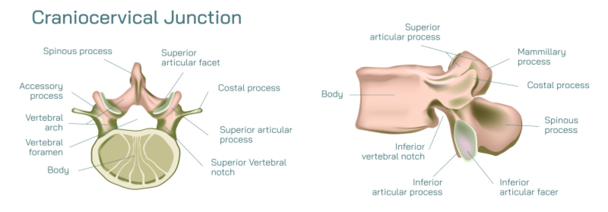
At the skeletal level, the craniocervical junction is formed by the occipital bone of the skull and the first two cervical vertebrae, known as the atlas (C1) and axis (C2). These three structures are unlike any others in the axial skeleton. The occipital bone features the foramen magnum, the large opening through which the brainstem descends into the spinal canal to become the cervical spinal cord. The atlas, ring-shaped and lacking a vertebral body, cradles the skull like a bony ring supporting a globe. It articulates with the occipital condyles to form the atlanto-occipital joint, which enables the nodding motion that allows the head to flex and extend. Beneath it lies the axis, distinguished by the dens (odontoid process) — a vertical projection that acts as a pivot for the atlas. Their connection, the atlanto-axial joint, permits the rotational movement of the head from side to side. Together, these two uppermost vertebrae provide a combination of mobility and stability unmatched elsewhere along the spine: the head can rotate, tilt, nod, and shift in multiple planes while still protecting the neural structures within.
Stability in the craniocervical region depends not only on bone architecture but on a sophisticated network of ligaments, which bind the skull, atlas, and axis tightly enough to prevent harmful displacement while preserving freedom of movement. The transverse ligament of the atlas is perhaps the most crucial, holding the dens securely against the anterior arch of the atlas so that motion does not compress the spinal cord. The alar ligaments, extending from the dens to the occipital bone, limit excessive rotation and lateral flexion of the head. The tectorial membrane, a continuation of the posterior longitudinal ligament inside the spinal canal, reinforces the foramen magnum from within. Surrounding ligaments and joint capsules contribute additional reinforcement. These structures create a layered, interlocking reinforcement system around the dens and the spinal canal. Because of their essential stabilizing role, ligament injury in this region poses serious risk, and even subtle ligamentous laxity can permit abnormal motion that threatens the brainstem or spinal cord. The careful balance between mobility and stability reflects evolutionary refinement: the early vertebrates that emerged from aquatic origins needed to lift and turn the head while still protecting the delicate neural circuits linking brain and body.
1. This area contains pathways essential for sustaining life — the neural networks regulating breathing, heartbeat, blood pressure, swallowing, and consciousness. A dense collection of ascending and descending nerve tracts runs through this narrow corridor. Among the most important are the corticospinal tracts, which deliver voluntary motor commands from the cerebral cortex to muscles throughout the body, and the spinothalamic and dorsal column pathways, which relay sensory information from the body back to the brain. At this level, many motor fibers have not yet crossed or are still crossing from one side of the body to the other, which is why injury near the craniocervical junction can produce profound neurological consequences far more severe than injuries in lower spinal segments.
Cranial nerves also play a prominent role in this anatomical region. Several cranial nerve nuclei are located inside the brainstem immediately above the foramen magnum, and many nerve roots pass through or near the craniocervical junction to exit the skull. The vagus nerve, which mediates autonomic control of the heart and digestive tract, descends alongside this region, while the accessory nerve, which innervates neck and shoulder muscles, emerges partly from the upper cervical cord. The phrenic nerve, responsible for diaphragm movement and breathing, receives fibers from the high cervical spinal cord segments (C3–C5). The integrity of the craniocervical junction therefore affects the most basic functions necessary for survival. Disruption here can alter vocal production, swallowing, eye movement, posture, respiratory control, and autonomic stability.
A further structural system within the craniocervical region is the dural membrane, the tough outer covering of the central nervous system. As it lines the skull and extends down the spinal canal, it encloses the cerebrospinal fluid (CSF) that cushions and protects neural tissue. The shape of the foramen magnum and the arrangement of the dura determine how CSF flows between the cranial cavity and spinal canal. Even subtle changes in alignment or anatomical configuration can affect this circulation, contributing to headaches, pressure imbalance, or neurological symptoms. Congenital variations such as Chiari malformation, in which part of the cerebellum extends downward toward the foramen magnum, illustrate how crowding in this space disrupts both CSF flow and neural signaling. Similarly, inflammatory diseases, tumors, or degenerative changes in the atlas–axis complex can impinge on the dura or alter intracranial pressure dynamics.
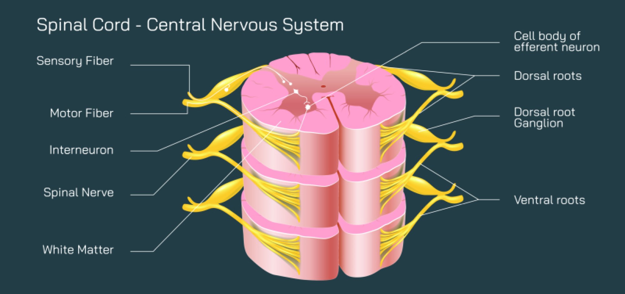
The spinal cord emerging from the craniocervical junction continues downward surrounded by vertebrae, ligaments, discs, and meninges, but its functional organization begins here. The C1 and C2 spinal segments convey motor and sensory information to and from the scalp, face, throat, and neck, while the higher spinal cord regulates communication with the entire body below. As the segments descend, the spinal cord gives rise to spinal nerve roots, each carrying mixed motor and sensory fibers. The spacing of spinal nerves and their dermatomal maps are rooted in embryonic development, but their architecture remains tightly constrained by the protective confines of the craniocervical canal. The high cervical cord also contains neural circuits responsible for proprioception, the internal sense of body position that allows balance and controlled movement. Through these pathways, the craniocervical junction integrates not only brain and body coordination but the sense of spatial orientation that allows humans to stand, walk, and navigate in three-dimensional environments.
2. Degenerative diseases may lead to instability or narrowing of the spinal canal. Inflammation or infection may affect ligaments or joint capsules, producing pain and neural irritation. Even small abnormalities can produce significant symptoms because the neural tissues in this area cannot tolerate compression or reduced blood supply. Symptoms affecting this region may range from neck pain, headaches, dizziness, and balance disturbances to tingling, muscle weakness, visual or swallowing problems, or more severe neurological impairment. Diagnosis often relies on magnetic resonance imaging, computed tomography, dynamic radiography, and neurological examination to evaluate bone structure, ligament integrity, neural tissue, and CSF flow.
Despite these risks, the craniocervical junction normally functions with remarkable reliability, allowing millions of people to move, think, breathe, and perceive effortlessly every day. The region exemplifies how anatomy enables physiology: the atlas and axis create an unparalleled range of head motion while protecting the most delicate information pathway in the body; ligaments function with surgical precision to prevent dangerous movement while permitting controlled articulation; the brainstem transitions into the spinal cord within a protected bony canal that supports rather than obstructs neural conduction; and the dura and cerebrospinal fluid cushion the neural axis against mechanical stress. The coordination of all these components permits the human head to serve as a sensory hub, cognitive center, and mechanical interface without compromising the continuity of neural commands and sensations.
Ultimately, the craniocervical junction stands as one of the finest examples of structural and functional integration in human biology. It embodies the transformation from cranial to spinal organization, supporting both the motion of the head and the transmission of signals that animate every part of the body. It reveals how evolution refined not only bones and joints for mechanical efficiency but also neural passageways for survival-critical communication. To understand the craniocervical junction is to recognize the extraordinary relationship between skeletal engineering and neural design — a partnership that enables mobility, sensation, coordination, cognition, and life itself.