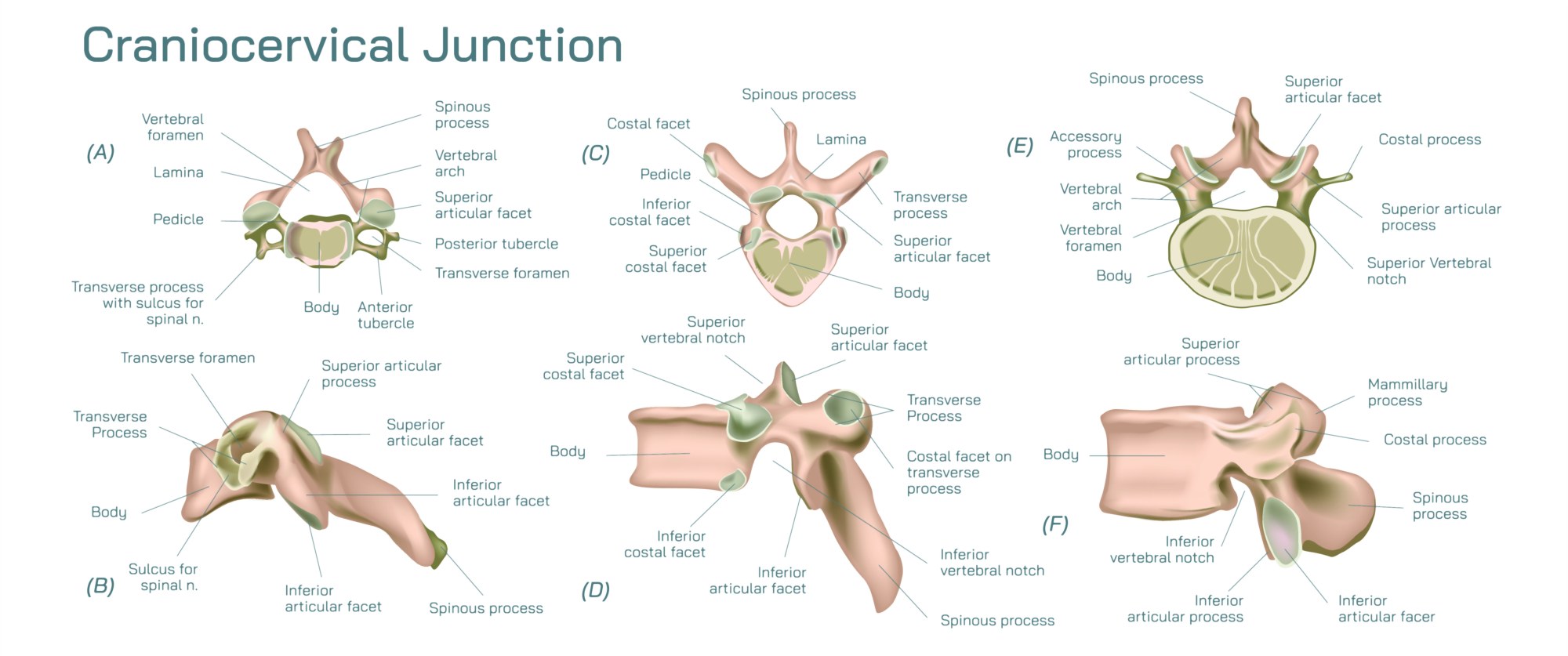
Detailed Overview of the Craniocervical Junction and Its Structural Importance
The craniocervical junction represents one of the most intricate anatomical regions in the human body. It forms the connection between the skull and the upper cervical spine, creating a structural bridge that allows controlled motion while maintaining safe passage for neural elements. This region includes the base of the skull, the first cervical vertebra known as the atlas, and the second vertebra known as the axis. These components create a coordinated system designed to support head movement, protect neural tissue, and stabilize posture.
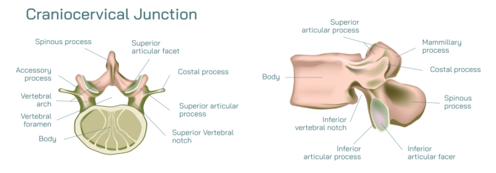
At the uppermost point of the junction lies the occipital base, a curved bony area that rests upon the atlas. This connection forms the atlanto-occipital joint, a structure that permits nodding and subtle front-back motion. The smooth articulation between these surfaces ensures controlled flexion and extension without strain. Beneath this joint sits the atlas, a ring-shaped bone designed without a typical vertebral body. Instead, it forms a wide arch that accommodates the spinal canal while distributing weight from the head.
Directly below the atlas lies the axis, the vertebra distinguished by its projecting odontoid process. This structure provides a pivot for rotation, enabling the head to turn from side to side. The interaction between the atlas and axis forms the atlanto-axial joint, a key driver of upper-neck rotation. This joint allows a significant portion of head movement while maintaining stability through its ligamentous support. The ligaments surrounding this region play critical roles in restricting excessive motion that could compromise neural pathways.
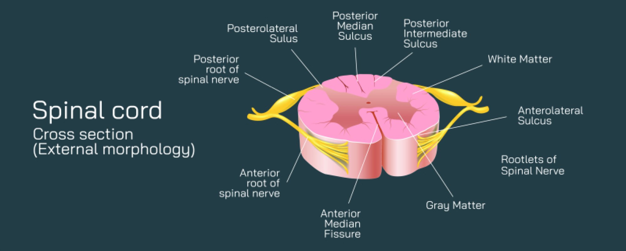
One of the essential features of the craniocervical junction is its relationship with the spinal canal. The transition from the brainstem to the spinal cord occurs within this area, making its alignment crucial for neural function. Any disruption in the spacing, curvature, or structural balance can affect signals passing through the canal. This region also houses vital blood vessels that supply the brain and upper spinal cord, increasing the importance of its mechanical stability.
Externally, the craniocervical junction shows a balance of supportive and mobile structures. Muscle groups attach around the bone surfaces, creating a system that guides coordinated movement. These muscles assist in stabilizing the head, controlling rotation, and supporting posture. Their combined action ensures the joint surfaces remain aligned during daily activity. The bony margins offer strong attachment points for these muscles, allowing efficient force transfer.
Ligaments in the region further reinforce stability. The transverse ligament holds the atlas in proper relationship with the axis, preventing harmful displacement. Additional connective structures support the occipital base, ensuring that the skull does not shift excessively on the cervical arch. These ligaments maintain tension that keeps the region secure even during dynamic motion, offering continuous protection for neural structures passing through the canal.
Understanding the craniocervical junction requires appreciating its dual purpose of mobility and protection. It enables head flexion, rotation, and positioning while shielding the neural elements that descend from the brain. The combined influence of bone shape, joint design, ligament tension, and muscle support creates a balanced system that responds smoothly to motion demands. Disturbances in this region, whether structural or functional, can affect coordination, motion control, and neural transmission.
Through its layered organization, the craniocervical junction acts as both a mechanical support system and a protective frame. Its form and function together create a structural gateway that guides movement, supports sensory input, and preserves neural continuity. This detailed understanding helps illustrate why this region is essential for balance, posture, and coordinated head-neck interaction.