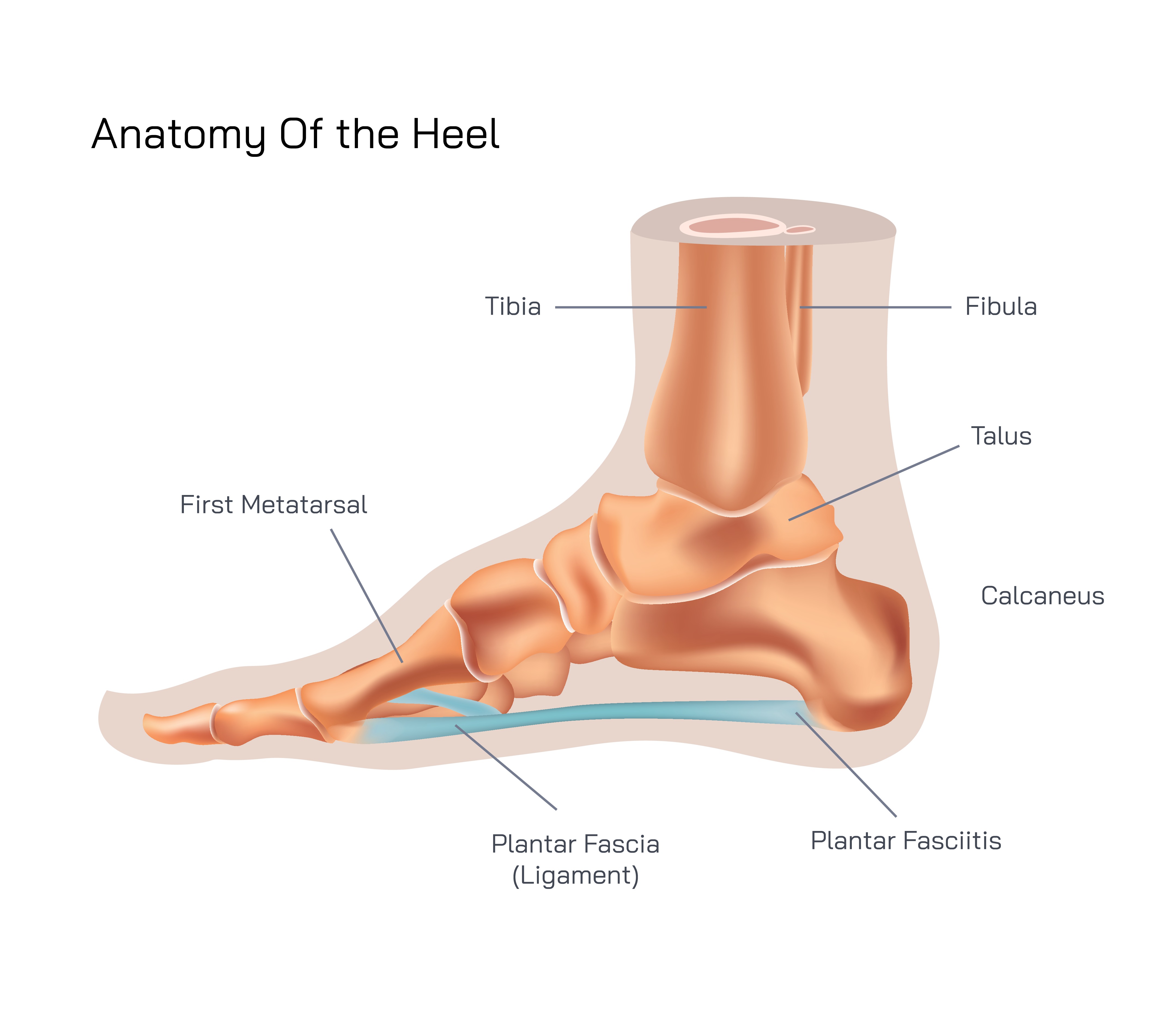
The anatomy of the heel represents one of the most vital structural pillars of the human body, serving as the foundation for movement, body weight support, balance, and shock absorption. Although the heel may appear simple at first glance, its internal composition reveals a sophisticated system of bones, connective tissues, muscles, blood vessels, and nerves working together with mechanical precision. Every step taken begins with the heel’s strategic contact with the ground, and this moment of impact shapes the entire gait cycle. The design of the heel ensures that immense pressure from body mass and ground reaction force is dispersed safely rather than concentrated on delicate upper joints, protecting the ankles, knees, hips, and spine from injury. Without its unique structure, human locomotion would be extremely unstable and painful.
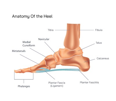
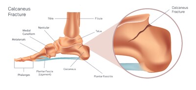
At the core of the heel lies the calcaneus bone, the largest bone in the foot and the fulcrum that receives the initial impact during walking and running. This dense bone connects superiorly with the talus to form the subtalar joint, a highly specialized joint that enables the foot to adapt to uneven surfaces by producing inversion and eversion movements. The back of the calcaneus serves as the strong anchor for the Achilles tendon, the thickest tendon in the human body, which transmits force from the calf muscles to generate powerful push-off actions during locomotion. The lower surface of the calcaneus houses the heel pad, a uniquely engineered fat-filled cushion formed of fibrous septa that preserve their shape despite constant compression. This heel pad plays a decisive role in absorbing shock so that the skeleton above does not experience repeated trauma from daily movements.
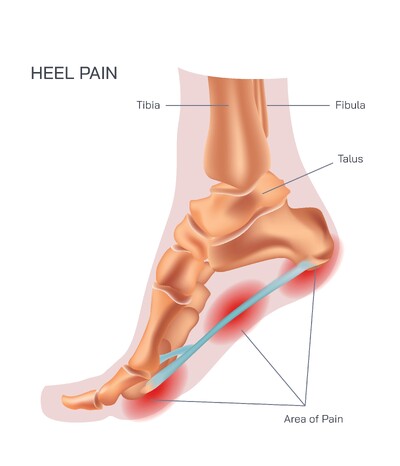
The heel is structurally inseparable from the plantar fascia, one of the most critical connective tissues of the foot. Running from the calcaneus to the toes, the plantar fascia acts like a tensioned bowstring that supports the medial longitudinal arch of the foot and prevents the arches from collapsing under pressure. When properly aligned and elastic, the structure of the plantar fascia allows the heel to convert downward pressure into forward propulsion, making motion smoother and more energy efficient. However, strain or microtear within this structure can result in severe heel pain known as plantar fasciitis, a common orthopedic condition triggered by overuse, improper footwear, or disrupted biomechanics.
Muscles and tendons surrounding the heel add dynamic control, allowing both gross movement and fine balance correction. Extrinsic muscles originating from the lower leg, including the gastrocnemius and soleus, transmit force through the Achilles tendon to power walking and running. Meanwhile, intrinsic muscles housed within the foot contribute to the alignment of the toes and balance of the arches. Their coordinated action ensures that the heel does not merely bear weight passively but adapts responsively to every terrain, speed, and posture change. Ligaments around the heel reinforce joint stability, preventing excessive motion that could lead to sprains. Together, these tissues maintain a complex but harmonious balance between rigidity and flexibility.
The heel also contains an intricate network of sensory nerves that play a subtle but essential role in movement. These nerves continually monitor pressure changes, foot temperature, surface texture, and balance, transmitting real-time information to the brain. The body responds by adjusting posture and muscle activation patterns almost instantaneously, preventing falls and enabling stable movement without conscious effort. Blood vessels woven through the heel supply oxygen and nutrients to muscles and connective tissues while supporting the healing of microscopic damage that inevitably occurs during repeated motion. Any disruption in circulation or nerve integrity can affect not only heel comfort but also overall gait efficiency.
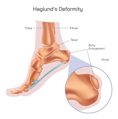
Understanding the anatomy of the heel extends beyond academic curiosity; it carries practical implications for healthcare, sports performance, rehabilitation, and everyday lifestyle. When heel mechanics become impaired—whether from overuse injuries, poor footwear, excessive standing, aging, obesity, trauma, or structural imbalances—mobility can decline dramatically. Pain conditions such as Achilles tendonitis, plantar fasciitis, heel spurs, bursitis, or nerve compression can profoundly affect daily functioning and quality of life. Conversely, supporting heel anatomy through proper alignment, footwear with adequate cushioning and arch support, flexibility training, and strength conditioning can protect the heel and ensure efficient motion throughout life.
The heel is ultimately a biological masterpiece designed to endure tremendous load while enabling graceful and coordinated movement. It is the unsung foundation that stabilizes every step, connects the lower limb to the ground, and protects the skeletal system from the repeated shocks of locomotion. Appreciating its anatomy allows us not only to understand how humans move but also to recognize the importance of maintaining foot health for long-term mobility and physical independence.