Human Knee Anatomy – Structure, Function, and Key Components
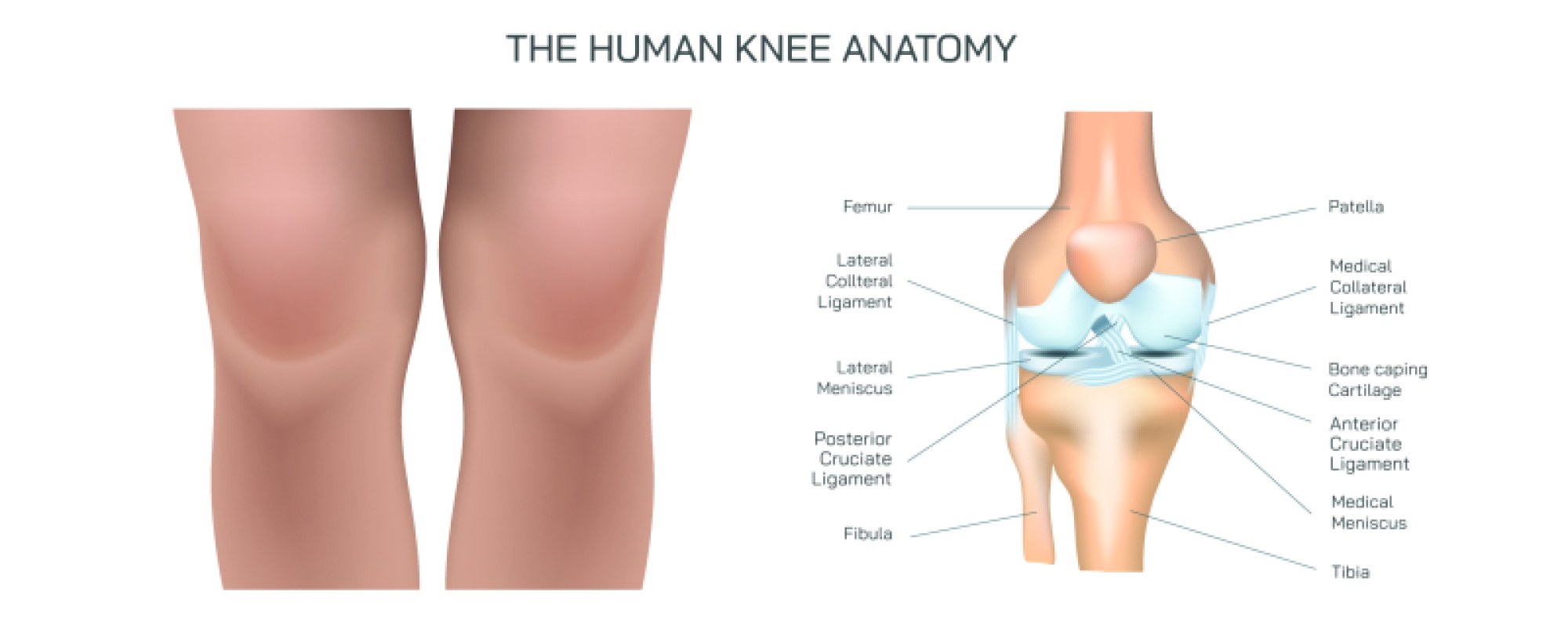
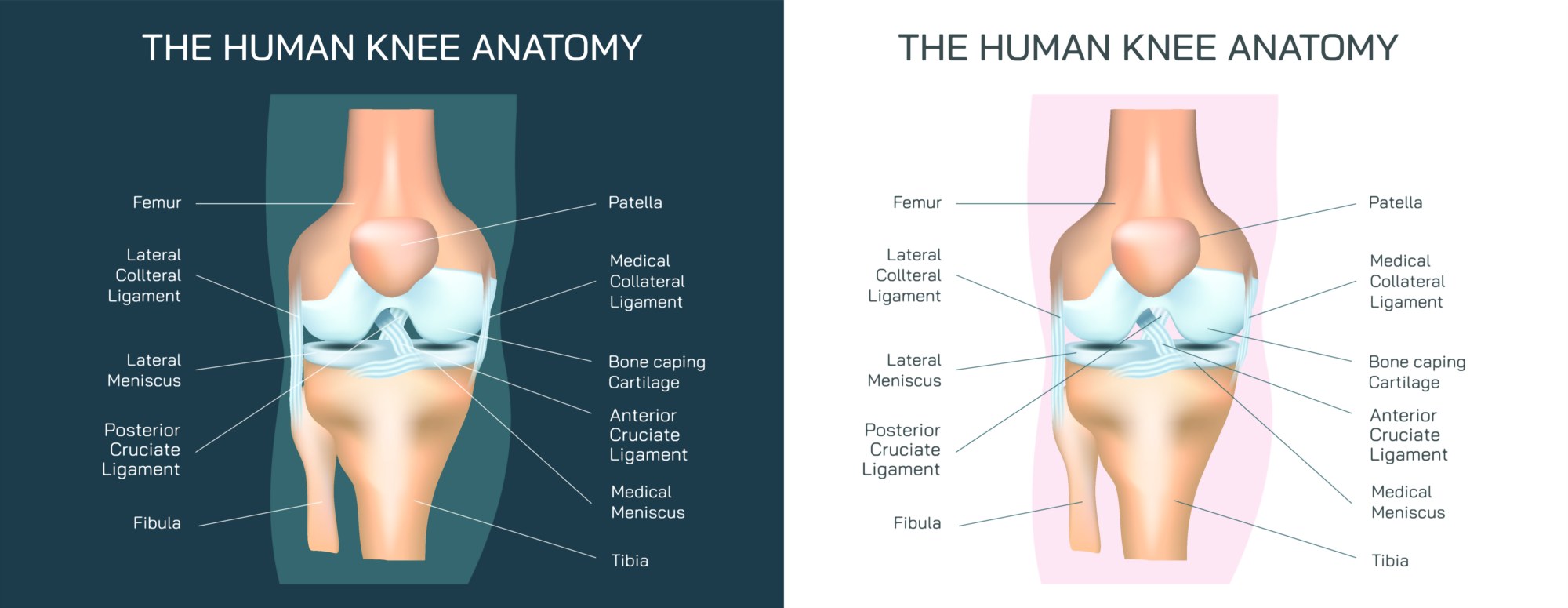
The human knee is one of the most complex and vital joints in the human body, responsible for movement, support, and stability. It is a hinge joint that connects the femur (thigh bone), the tibia (shin bone), and the patella (kneecap), allowing for flexion, extension, and slight rotational movement. The knee supports the weight of the body during activities like walking, running, jumping, and squatting, making it highly susceptible to injuries and degeneration.
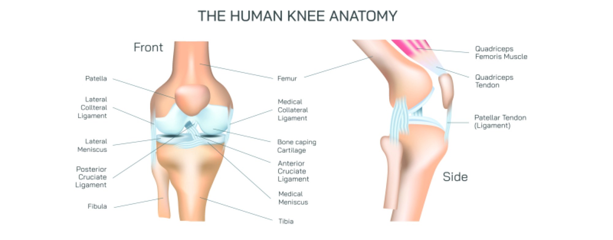
Bones of the Knee: The knee joint primarily involves three bones. The femur forms the upper portion, the tibia forms the lower portion, and the patella sits anteriorly, protecting the joint and aiding in the leverage of the quadriceps tendon. The articulation between these bones is cushioned by articular cartilage, which reduces friction and distributes mechanical stress. The menisci – two C-shaped cartilaginous structures (medial and lateral) – act as shock absorbers and help stabilize the joint during motion.
Ligaments of the Knee: The knee relies on strong ligaments for stability. The anterior cruciate ligament (ACL) and posterior cruciate ligament (PCL) control forward and backward motion of the tibia relative to the femur. The medial collateral ligament (MCL) and lateral collateral ligament (LCL) provide side-to-side stability, preventing excessive valgus and varus movements. These ligaments work together to maintain joint integrity during dynamic activities.
Muscles and Tendons: The knee joint is supported by surrounding muscles and tendons. The quadriceps in the front of the thigh extend the knee, while the hamstrings at the back enable flexion. The patellar tendon connects the patella to the tibia, and the quadriceps tendon connects the quadriceps muscles to the patella. Coordinated contraction of these muscles ensures smooth, controlled movement while maintaining joint stability.
Joint Capsule and Synovial Membrane: The knee is enclosed in a joint capsule lined with a synovial membrane that secretes synovial fluid. This fluid lubricates the joint, nourishes cartilage, and reduces friction between articulating surfaces, enabling pain-free movement.
Function and Biomechanics: The knee’s primary functions include weight-bearing, mobility, and shock absorption. Its unique hinge structure allows flexion (bending) and extension (straightening), with limited rotation when the knee is flexed. Proper alignment of bones, ligaments, and muscles ensures efficient energy transfer, stability, and balance during movement.
Clinical Significance: The knee is prone to injuries such as ligament tears, meniscus damage, and cartilage degeneration. Understanding its anatomy is critical for orthopedic diagnosis, physical therapy, sports medicine, and surgical procedures like knee arthroscopy or joint replacement. Detailed anatomical knowledge aids in injury prevention, rehabilitation, and improving joint function.
In conclusion, the human knee is a highly intricate joint that integrates bones, ligaments, muscles, cartilage, and synovial structures. Its complex biomechanics support mobility, stability, and weight-bearing functions. Comprehensive understanding of knee anatomy is essential for medical professionals, students, athletes, and anyone interested in human physiology or musculoskeletal health. This knowledge not only aids in treatment and rehabilitation but also enhances awareness of how to maintain healthy, functional knees throughout life.