Femur Bone Anatomy Showing Structure, Regions, and Functions of the Longest Human Bone
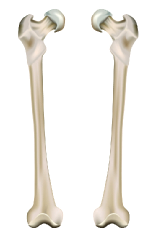
The femur is the largest, longest, and strongest bone in the human body, forming the primary structural support of the thigh and serving as a central component of posture, balance, and movement. A vector illustration highlighting the anatomy of the femur—its structure, major regions, and physical and physiological functions—offers a clear visual map of why this bone is considered the foundation of human locomotion. Even though most people recognize the femur merely as the “thigh bone,” its anatomical complexity reveals a highly specialized design optimized for weight-bearing, shock absorption, muscle leverage, stability of the hip and knee joints, and protection of the underlying neurovascular systems. When the femur is studied in detail from top to bottom, it becomes clear that its shape is not arbitrary; each region, contour, and landmark contributes to efficient, powerful motion and to the transfer of forces during everyday movement.
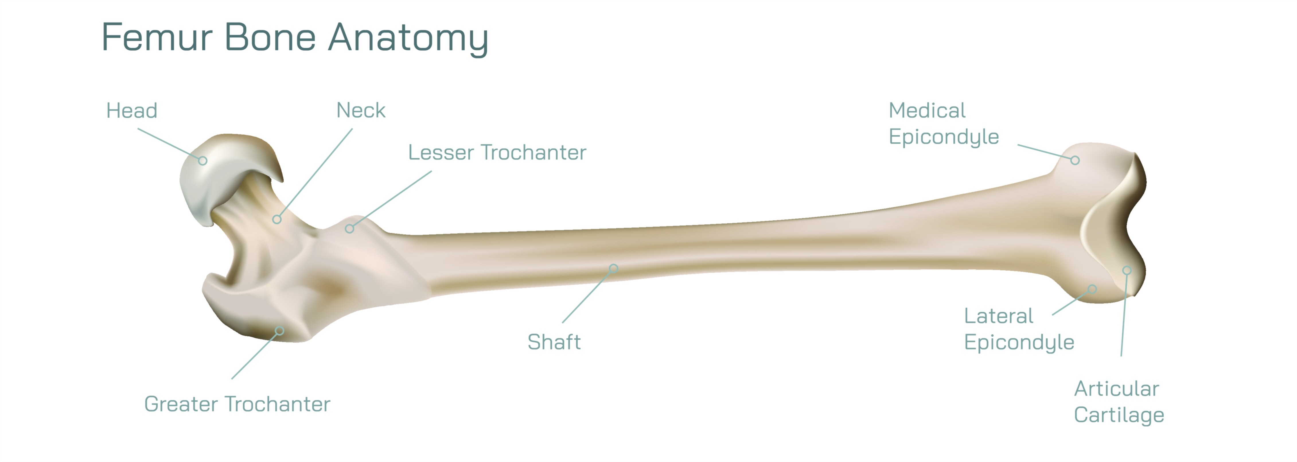
The proximal end of the femur begins with the femoral head, a smooth, rounded, spherical structure that fits securely into the acetabulum of the pelvis, forming the hip joint. This ball-and-socket connection allows for multidirectional motion—flexion, extension, rotation, abduction, and adduction—making the hip one of the most mobile joints in the skeleton while still supporting the body’s full weight. The head of the femur is covered with thick articular cartilage to reduce friction and absorb force across the joint. Just below it lies the femoral neck, a narrowed bridge that angles the head toward the long shaft. Although this region allows freedom of hip movement, it is also biomechanically vulnerable and frequently involved in fractures among older adults. Extending from the neck are two prominent bony projections: the greater trochanter on the lateral side and the lesser trochanter on the posteromedial side. These trochanters do not exist for aesthetic shape; they serve as powerful muscle attachment points for the gluteal muscles, iliopsoas, and other deep rotators that drive hip movement and stabilize the pelvis during standing and walking.
Below the trochanters, the femur transitions into the shaft, which forms the majority of the bone’s length. The shaft is gently bowed forward—a shape that increases resilience under vertical compression. It is composed of thick cortical (compact) bone, which is densely structured to withstand high axial loads, and internally lined with trabecular (spongy) bone that absorbs shock and disperses mechanical stress during dynamic motion. A vector illustration of the shaft often highlights the linea aspera, a ridge that runs down the posterior surface. This feature serves as the anchoring site for adductor muscles and quadriceps components and represents one of the most important structural reinforcements of the femoral design. The very presence of the linea aspera shows how intimately bone shape and muscle function are linked; every muscle that attaches to the femur influences gait, posture, knee stability, and power transmission.
Approaching the lower thigh, the femur broadens into the distal end, which forms the upper portion of the knee joint. Here, two large rounded prominences—the medial and lateral condyles—meet the tibia and articulate with the patella. These condyles distribute weight across the knee during walking, running, sitting, and jumping. Between them lies the intercondylar fossa, which accommodates the cruciate ligaments essential for knee stability. On the front surface of this region is the patellar surface, a smooth groove that guides the patella during flexion and extension. A detailed vector illustration showing the condyles, epicondyles, and patellar surface gives context to the biomechanical stresses that the knee experiences and explains how the femur forms the upper half of one of the most active joints in the body.
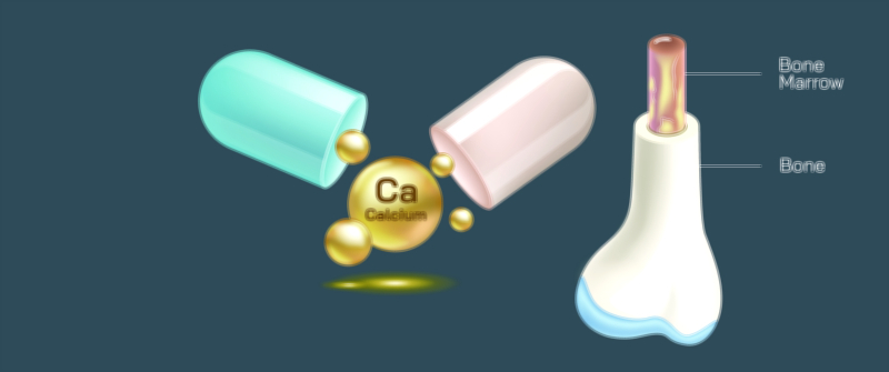
The internal composition of the femur is as vital as its external shape. The thick cortical wall provides strength for weight-bearing, but the bone marrow within the medullary cavity contributes to physiology that extends beyond movement. In children and young adults, red bone marrow fills much of this cavity, producing red blood cells, white blood cells, and platelets. With age, red marrow transitions gradually into yellow marrow, which stores lipids and can convert back to red marrow when blood cell production increases. A vector view of the femur cut cross-sectionally emphasizes that bone is not an inert pillar but a living tissue with ongoing metabolic and hematopoietic activity.
The functions of the femur expand far beyond its role as a rigid support structure. As the primary weight-bearing bone of the lower body, the femur transmits forces between the hip and knee every time a person stands upright. It absorbs and redistributes impact energy during motion, protects deep arteries, veins, and nerves that run alongside it, and provides leverage for muscles that enable powerful gait, sprinting, jumping, lifting, and balance. Through its internal remodeling system, the femur responds dynamically to life: when forces increase from physical training, the bone thickens; when forces decrease from immobilization or sedentary behavior, the bone gradually weakens. This adaptability is central to understanding how good posture and physical activity reinforce skeletal health across decades.
Each region of the femur plays a specific role in coordinated biomechanics:
• The femoral head and neck enable multidirectional hip motion.
• The trochanters anchor powerful muscles that lift and rotate the thigh.
• The shaft transmits body weight through a strong but flexible arc.
• The distal condyles form the hinge surface of the knee and support patellar movement.
• The bone marrow cavity sustains blood formation and stores energy.
Together, these structures allow the femur to perform two simultaneous tasks—maintaining rigid stability while facilitating fluid movement.
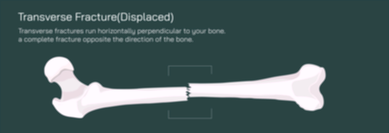
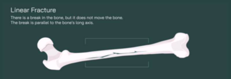
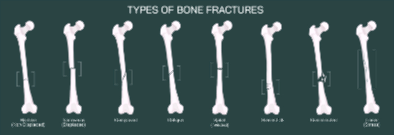
A vector illustration that combines the external regions, internal structure, muscle attachment points, and joint articulations creates a unified visual narrative of how the femur supports life. For learners, this kind of diagram transforms anatomy from a list of landmarks into an integrated system, showing why fractures at different femoral regions behave differently, why certain muscles produce specific hip and knee actions, and why bone density is so crucial for long-term mobility. For patients and families, such a visual helps make sense of injuries, joint replacements, and rehabilitation strategies. And for anyone studying the musculoskeletal system, it reinforces a simple reality: the femur is more than the longest bone of the body — it is a biomechanical masterpiece linking strength, movement, and physiology into one anatomical structure designed to carry the body through a lifetime of motion.