Human Knee Anatomy Illustration: Bones, Ligaments, and Joint Structure Explained
The human knee is one of the most complex and essential joints in the body, and an illustration that reveals its internal structure provides insight into how this remarkable joint supports movement, stability, and weight-bearing throughout daily life. Although the knee appears simple from the outside—functioning like a hinge that bends and straightens the leg—an anatomical cross-section highlights an intricate system of bones, ligaments, tendons, cartilage, and connective tissues working together to create strength and mobility. The knee endures enormous physical demands, supporting the body’s weight during standing, walking, running, jumping, kneeling, squatting, and sudden directional changes. To enable this range of actions while maintaining stability, its internal design balances rigid support with flexible cushioning and precise mechanical control.
The skeletal foundation of the knee comprises three primary bones that meet at the joint: the femur (thighbone), tibia (shinbone), and patella (kneecap). In an illustration, the femur descends into the joint through two rounded prominences called the medial and lateral femoral condyles, which glide across the flattened surfaces of the tibia known as the medial and lateral tibial plateaus. These articulating structures form the tibiofemoral joint—the main hinge component of the knee. The patella, a triangular sesamoid bone embedded within the quadriceps tendon, rests in a groove on the front of the femur called the trochlear groove, forming the patellofemoral joint. The patella acts like a fulcrum, improving the mechanical advantage of the quadriceps muscle and enhancing knee extension. In diagrams, it sits prominently over the joint, sliding vertically during motion to maintain alignment and distribute forces.
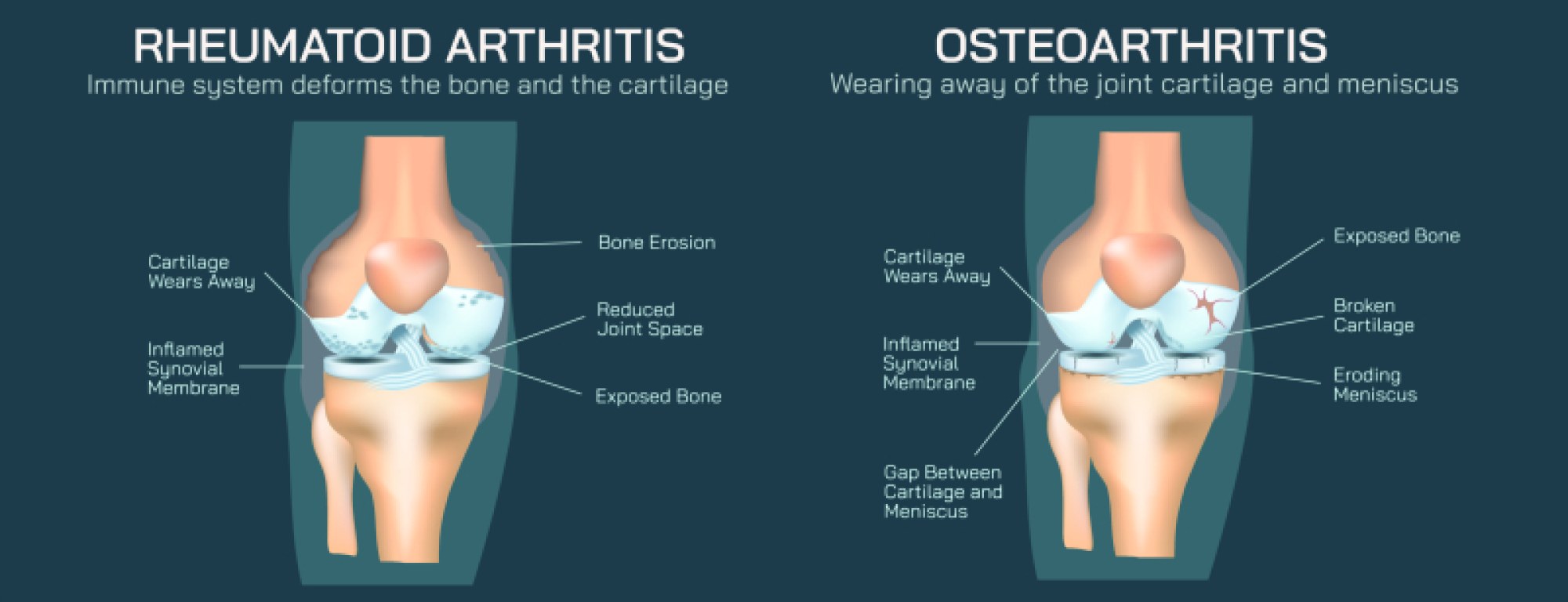
A central component of knee illustrations is the joint capsule, a fibrous envelope that encloses the entire knee structure. Within this capsule lies the synovial membrane, which produces synovial fluid, a lubricating substance that reduces friction and nourishes the articular cartilage that lines the bone surfaces. The smooth articular cartilage covering the femoral condyles, tibial plateaus, and posterior patella allows motion to occur with minimal resistance. Visual diagrams often show cartilage as a glossy, bluish-white layer, emphasizing the importance of low-friction gliding surfaces. When cartilage wears down—whether through age, repetitive stress, or injury—the resulting bone-on-bone contact leads to osteoarthritis, pain, swelling, and restricted movement. Anatomical illustrations frequently highlight this degeneration to help explain joint pathology.
At the core of knee stability are the four primary ligaments, typically emphasized in bold lines and clean labels in medical diagrams because of their crucial mechanical roles. The anterior cruciate ligament (ACL) and posterior cruciate ligament (PCL) form a crossing “X” structure in the center of the joint. The ACL runs from the anterior tibia to the posterior femur and prevents forward shifting of the tibia and excessive rotational movement. The PCL runs from the posterior tibia to the anterior femur and prevents backward displacement of the tibia. Injuries to the ACL are common in sports, particularly during sudden pivoting motions, while PCL injuries more often occur from direct trauma. Illustrations that cut through the joint typically show the ACL angled forward and the PCL angled backward to visualize their complementary stabilizing effect.
Flanking the interior cruciate ligaments are two major stabilizers on the outer surfaces of the joint: the medial collateral ligament (MCL) along the inner side of the knee and the lateral collateral ligament (LCL) along the outer side. The MCL prevents the knee from buckling inward (valgus stress), while the LCL prevents outward buckling (varus stress). In anatomical diagrams, the MCL appears as a broad, flat band merging seamlessly with the medial meniscus, while the LCL appears as a thinner, cord-like band attached directly between the femur and fibula. These ligaments are vital for static stability during walking and standing, preventing unwanted lateral motion while allowing smooth flexion and extension.
Two distinct menisci—the medial meniscus and lateral meniscus—sit atop the tibial plateaus and are usually highlighted as crescent-shaped, fibrocartilaginous wedges in illustrations of the knee interior. Acting as shock absorbers and load distributors, these structures cushion the joint and enhance stability by deepening the tibial articular surfaces. Because the medial meniscus attaches more firmly to the MCL, it is less mobile and more prone to tearing during twisting motions. The lateral meniscus is more flexible and mobile, reducing its injury risk in many cases. Diagrams concerning meniscal tears often show jagged splits or flap-like displacements within the menisci, clarifying why such injuries can create painful clicking, locking, and swelling.
Musculature and tendons play an equally crucial role in knee mechanics, and detailed illustrations often include the quadriceps tendon above the patella and the patellar tendon (or patellar ligament) below it, anchoring the kneecap to the tibial tuberosity. The quadriceps muscles enable knee extension, while the hamstrings—crossing the posterior of the joint—assist in flexion. Although muscles are not bones or ligaments, most knee diagrams include their tendon attachments to show the integrated movement system. The iliotibial (IT) band along the lateral thigh is also commonly shown because it contributes to lateral support and is associated with overuse syndromes in athletes.
Inside the joint, bursae—small fluid-filled sacs—help reduce friction. The largest and most commonly illustrated is the prepatellar bursa, located in front of the patella. Additional bursae surround the joint capsule to cushion tendons and ligaments during motion. When inflamed, these fluid sacs swell and cause localized pain, a condition represented in illustrations by enlarged or fluid-filled pouches that restrict normal motion.
Blood supply and innervation diagrams highlight the vascular networks and nerves that maintain knee health and function. The popliteal artery, running behind the knee, branches into vessels that nourish bone, cartilage, and surrounding tissues. Nerves such as the tibial nerve and common fibular (peroneal) nerve supply sensation and muscular control. These structures are sometimes included in educational illustrations to reinforce the knee’s sensitivity to trauma and why injuries may cause pain, numbness, or muscle dysfunction.
Because the knee endures constant mechanical demand, injury diagrams are often paired with anatomical illustrations to explain conditions such as ligament tears, meniscus injuries, tendonitis, bursitis, dislocation of the patella, and degenerative joint disease. Images comparing healthy and damaged structures highlight how swelling, misalignment, and tissue breakdown alter movement and stability. These comparisons emphasize that even a single compromised structure may disrupt the function of the entire joint.
Ultimately, an illustration of the human knee goes far beyond identifying bones and ligaments—it reveals the coordinated design of a joint that must be both strong and flexible, both stable and mobile. It shows how the femur, tibia, and patella interlock to create a foundation for weight-bearing; how ligaments and menisci prevent shifting, slipping, and compression damage; and how cartilage, synovial fluid, and cushioning structures protect the joint from friction and shock. By visualizing these elements together, the anatomy illustration conveys that the knee is not merely a hinge—it is a sophisticated biological mechanism supporting nearly every form of human movement, from the smallest step to the most powerful athletic motion, and its structural harmony is crucial to lifelong mobility and quality of life.