Healthy Knees Vector Illustration Showing Strong Joints, Mobility, and Cartilage Support for Active Movement
Healthy knees lie at the center of nearly every major movement the human body performs—walking, running, jumping, climbing stairs, sitting down, standing up, and bearing weight throughout the entire lifespan. Even though the knee is often thought of as a simple hinge, its true structure is much more complex, combining bone, cartilage, ligaments, tendons, muscles, and synovial fluid into a coordinated biomechanical system that absorbs shock, stabilizes motion, and transfers force efficiently between the hips and the ankles. A vector illustration designed to show healthy knees, strong joints, mobility, and cartilage support provides an accessible visual map of what is happening beneath the skin each time we move. It transforms components that rarely receive conscious attention into a clear demonstration of how joint health enables an active lifestyle. Such an image is not only helpful in medical education but in sports science, physical therapy, orthopedic learning, and wellness contexts, because it allows people to visualize how properly functioning knees maintain strength and mobility while resisting the stresses of daily activity.
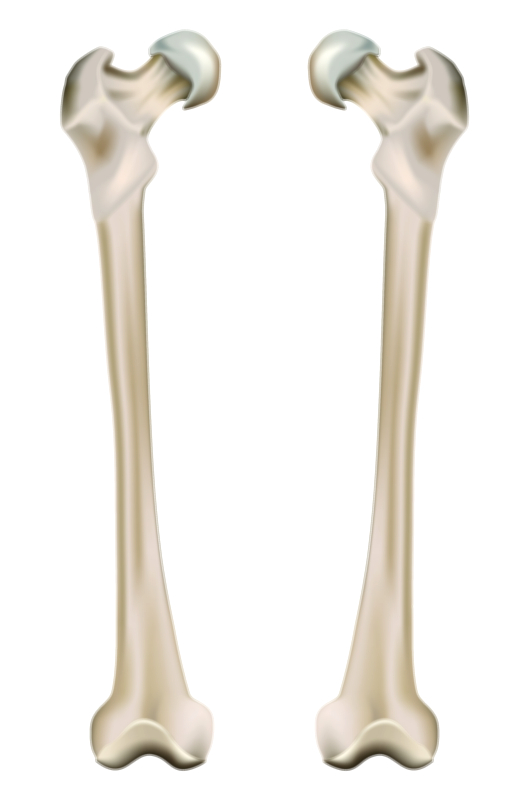
A healthy knee begins with solid bone alignment. The femur (thigh bone), tibia (shin bone), fibula, and patella (kneecap) must articulate smoothly in a configuration that distributes load evenly. The shape of the femoral condyles and the tibial plateau allows flexion and extension while permitting slight rotation when turning or changing direction. When bones align correctly, motion is fluid and strain is minimized. This is why a vector illustration often depicts not just the individual bones, but how their contact surfaces sync together under weight-bearing conditions. Proper alignment ensures that pressure does not concentrate on a single portion of the joint, which protects tissues from premature wear, pain, and injury.
The key to this smooth, gliding movement is cartilage—specifically, articular cartilage, a glossy cushioning layer that coats the bone ends within the joint. In a healthy knee, cartilage is thick, hydrated, and elastic, absorbing shock from steps, landings, and running strides. It reduces friction so dramatically that two cartilage surfaces sliding against each other generate less resistance than ice moving across ice. A vector illustration that highlights this cartilage layer emphasizes why its preservation is vital for lifelong joint mobility. Beneath the cartilage lies subchondral bone, which provides the supportive foundation needed to distribute load evenly. Together, they behave like a sophisticated shock-absorption system, allowing the knee to withstand repeated mechanical impact without discomfort.
Surrounding the knee joint is the synovial membrane, which produces synovial fluid—the knee’s internal lubricant. When represented visually, synovial fluid is often shown surrounding the cartilage surfaces in a thin layer that keeps them nourished and smooth. This fluid reduces wear and also transports nutrients to the cartilage, which has no blood supply of its own. Hydrated cartilage plus rich synovial fluid is a hallmark of a healthy knee and one of the primary reasons why movement feels effortless when joint tissues are in their optimal state.
Stabilization, meanwhile, comes from the remarkable ligament system. The anterior cruciate ligament (ACL) and posterior cruciate ligament (PCL) control forward and backward movement of the tibia relative to the femur. The medial and lateral collateral ligaments (MCL and LCL) protect the joint from collapsing inward or outward. When a vector illustration highlights these ligaments, it reinforces how each contributes to resisting excessive forces and preventing instability during activities like pivoting or landing from a jump. Strong ligaments work in synergy with surrounding tendons—especially the patellar tendon and quadriceps tendon—linking muscles to bones and powering knee extension. A balanced image of the knee often shows these connective tissues working together like ropes and pulleys to move the leg efficiently and safely.
Healthy knees also rely heavily on muscular support. Quadriceps stabilize the patella and help absorb shock before it even reaches the joint. Hamstrings protect the posterior structures and assist in controlling deceleration. Gluteal muscles support rotational stability and prevent inward knee collapse during movement. Calf muscles contribute to propulsion and shock reduction during running and landing. A vector illustration that includes these muscular connections demonstrates that joint health is not isolated—it is reinforced by full-body kinetic relationships. When surrounding muscles are strong and flexible, the knee does not carry stress alone, allowing cartilage and ligaments to remain protected over time.
At the center of joint balance are the menisci, two C-shaped fibrocartilage cushions that sit between the femur and tibia. They deepen the contact area between bones, distribute body weight evenly, reduce friction, and assist with stability during turning movements. Because they are vulnerable to strain from twisting injuries yet essential for joint longevity, an educational illustration of the knee often highlights their shape and load-bearing role. When meniscal cushioning is intact, the risk of cartilage damage and osteoarthritis decreases significantly, making their visual representation crucial in learning about knee mechanics.
A knee that functions properly supports more than mobility—it supports comfort, athletic performance, and confidence. People rarely recognize how dependent everyday life is on healthy joints until stiffness or pain interrupts movement. A vector illustration showing smooth articulation, hydrated cartilage, and stable ligaments helps people understand why knees feel different when things go wrong. It provides a baseline for comparison when learning about knee conditions such as osteoarthritis, meniscal degeneration, patellar misalignment, or ACL injuries. The contrast between a healthy knee and a deteriorating knee gives viewers a more intuitive understanding of how to protect their joints across the lifespan.
Prevention and maintenance also find clear expression in visuals. Hydrated cartilage relies on movement to circulate synovial fluid, making regular physical activity essential for joint nourishment. Strength training builds muscle support that reduces stress on ligaments and bone surfaces. Good posture and balanced gait protect cartilage from uneven wear. Weight management reduces load that the knees must carry. Stretching maintains flexibility in the quadriceps, hamstrings, and calves, allowing the patella to track properly and preventing unnecessary strain. A vector illustration can depict these relationships by showing improved cartilage hydration, greater load distribution, or reduced joint compression when the body is conditioned well.
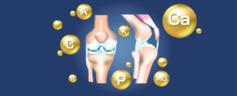
Nutrition also contributes to knee health, though it plays a supportive rather than mechanical role. Nutrients like collagen, vitamin C, vitamin D, omega-3 fatty acids, and minerals that strengthen bones indirectly help maintain the tissues around the knee. Educational artwork that places nutrition symbols around a knee diagram encourages the understanding that joint wellness is both biomechanical and metabolic—not purely structural.
Healthy knees contribute immensely to independence, happiness, and quality of life. The ability to move freely underlies everything from sports to daily routines to social engagement. When educational materials visualize strong knees with intact cartilage, aligned bones, smooth joint movement, and supportive tissues, they make the connection between invisible anatomy and lived experience clear. They remind viewers that maintaining knee strength does not simply prevent injury—it preserves energy, motivation, and confidence to stay active.
In its fullest form, a healthy knees vector illustration showing strong joints, mobility, and cartilage support becomes more than a diagram. It becomes a roadmap of human movement—showing how bones, cartilage, ligaments, muscles, and synovial fluid operate in harmony to carry the body with power and grace. It encourages learners, athletes, clinicians, and everyday individuals to respect the complexity of their joints and take proactive steps to support them. By turning anatomy into an understandable visual narrative, the illustration reinforces a message that everyone benefits from remembering: healthy knees are a foundation of lifelong motion, and caring for them today ensures mobility and vitality in the years to come.