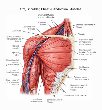
The anterior shoulder anatomy represents one of the most complex and functionally important regions of the human musculoskeletal system. It forms the structural foundation that allows the upper limb to move freely while maintaining stability and strength. The shoulder’s anterior view highlights key bones, joints, surfaces, and anatomical landmarks that work together to support a wide range of arm movements. Understanding this anatomy is essential for learning human biology, clinical medicine, physiotherapy, and biomechanics.
At the core of anterior shoulder anatomy are three major bones: the scapula, clavicle, and humerus. These bones connect the upper limb to the axial skeleton and create multiple articulations that allow flexibility and control. Unlike joints designed mainly for stability, the shoulder prioritizes mobility, which makes its anatomical organization both remarkable and vulnerable to injury.
The clavicle, commonly known as the collarbone, is a long, slender bone positioned horizontally at the front of the shoulder. In the anterior view, the clavicle extends from the sternum at its medial end to the acromion of the scapula at its lateral end. The sternal extremity of the clavicle articulates with the sternum, forming a stable connection between the upper limb and the central skeleton. This bone acts as a supportive strut that holds the shoulder away from the chest, allowing greater range of arm movement.
The acromioclavicular joint is formed where the lateral end of the clavicle meets the acromion of the scapula. This joint allows subtle movements that help the shoulder adapt during lifting and reaching. Though small, it plays an important role in transmitting forces from the upper limb to the trunk. Damage or misalignment in this joint can significantly affect shoulder function.
The scapula, or shoulder blade, is a flat, triangular bone that lies against the back of the rib cage but is clearly visible in anterior anatomical illustrations. The anterior surface of the scapula contains several important features. One of the most prominent is the subscapular fossa, a broad, shallow depression that occupies most of the anterior surface. This region provides attachment for muscles that stabilize and rotate the shoulder joint.
The coracoid process is a hook-like projection extending forward from the scapula. In anterior shoulder anatomy, it serves as a critical attachment point for muscles and ligaments that support shoulder stability. Its forward orientation allows it to act as a protective and anchoring structure, contributing to the overall strength of the shoulder girdle.
Another key structure visible from the anterior view is the glenoid fossa. This shallow socket is located on the lateral aspect of the scapula and forms the articulating surface for the head of the humerus. The shape of the glenoid fossa allows for a wide range of motion while relying on surrounding muscles and ligaments for stability. This design explains both the remarkable mobility of the shoulder and its susceptibility to dislocation.
The humerus is the long bone of the upper arm and plays a central role in shoulder movement. The head of the humerus is smooth and rounded, fitting into the glenoid fossa to form the glenohumeral joint. This joint is the main articulation of the shoulder and allows movements such as flexion, extension, rotation, and circumduction. The humeral head’s shape facilitates smooth movement while maintaining joint congruence.
Just below the humeral head are the greater and lesser tubercles. These bony prominences serve as attachment points for muscles that control shoulder rotation and stabilization. Their position influences muscle leverage and joint mechanics, making them essential landmarks in anatomical study and clinical assessment.
The scapular neck connects the glenoid fossa to the main body of the scapula. This narrowed region supports the joint surface and transmits forces generated during arm movement. Along the edges of the scapula, the medial and lateral borders define the shape and orientation of the bone. The inferior angle marks the lowest point of the scapula and serves as an important reference point for movement and muscle attachment.
From a functional perspective, the anterior shoulder anatomy allows the upper limb to operate efficiently in daily activities. Reaching forward, lifting objects, pushing, pulling, and throwing all depend on the precise alignment and coordination of these bones and joints. The clavicle maintains proper shoulder width, the scapula provides a mobile platform, and the humerus acts as a lever for movement.
Clinically, knowledge of anterior shoulder anatomy is essential for diagnosing injuries, planning surgical procedures, and designing rehabilitation programs. Fractures of the clavicle, dislocations of the glenohumeral joint, and damage to bony landmarks can all disrupt normal shoulder mechanics. Accurate understanding of anatomical relationships helps healthcare professionals restore function and reduce long-term complications.
In educational settings, anterior shoulder anatomy serves as a foundational topic for learning upper limb structure. The clear identification of bones, joints, and surfaces allows students to understand how structure supports function. Visual diagrams, like labeled anterior views, are especially valuable for grasping spatial relationships within the shoulder region.
The anterior shoulder anatomy reveals a finely balanced structural system that enables the upper limb to perform an extraordinary range of movements while still maintaining sufficient stability to support load-bearing and repetitive actions. This region is a key junction between the axial skeleton and the appendicular skeleton, and its design reflects the evolutionary priority placed on mobility, reach, and manual function in humans. Every bony surface, contour, and articulation visible from the anterior view contributes to the shoulder’s ability to combine freedom of motion with controlled strength.
One of the defining features of the anterior shoulder is the way the clavicle functions as a mechanical bridge. Positioned at the front of the shoulder girdle, the clavicle maintains the lateral position of the scapula and upper limb. Without this bone acting as a strut, the shoulder would collapse inward toward the chest, severely limiting arm movement. The clavicle transmits forces generated by the upper limb to the sternum, allowing loads to be distributed across the trunk rather than concentrated solely at the shoulder joint. Its slightly curved shape helps absorb stress and protect deeper structures during impact or heavy lifting.
The sternoclavicular articulation, though often overlooked, is the only true bony connection between the upper limb and the axial skeleton. From an anterior perspective, this joint anchors the shoulder girdle to the torso while still allowing subtle gliding and rotational movements. These small motions are essential for full elevation of the arm, as they permit the clavicle to move in coordination with the scapula during overhead activities.
The scapula’s anterior surface plays a major role in shoulder mechanics despite being less visible in everyday observation. The subscapular fossa dominates this surface and provides a broad area for muscular attachment that contributes to joint stability. The smooth, concave shape of this fossa allows muscles to lie close to the bone, optimizing force transmission without adding unnecessary bulk. This arrangement supports controlled rotation of the humerus and helps maintain alignment of the joint during movement.
The coracoid process is a particularly important landmark in anterior shoulder anatomy. Its forward-projecting position allows it to serve as a central anchoring point for several stabilizing structures. Because it extends anteriorly, it helps resist forces that would otherwise pull the shoulder joint forward. This is especially important during pushing movements or when the arm is raised and loaded. The coracoid process also provides a protective role, helping shield deeper neurovascular structures that pass through the shoulder region.
The glenoid fossa represents a unique compromise between mobility and stability. Unlike deep socket joints designed primarily for weight-bearing, the glenoid is shallow, allowing the humeral head to move freely in multiple directions. From the anterior view, the orientation of the glenoid fossa influences how the arm naturally rests and moves forward. The surrounding bony margins and connective tissues enhance joint congruence without sacrificing motion. This design allows the shoulder to perform complex actions such as reaching, throwing, and lifting with precision.
The humerus is the principal lever of the upper limb, and its proximal anatomy is critical to shoulder function. The spherical shape of the humeral head allows smooth articulation with the glenoid fossa. Its orientation facilitates forward elevation and rotation, which are especially evident in anterior movements like lifting an object in front of the body. The transition from the head to the shaft distributes mechanical stress and reduces the risk of localized strain during repeated motion.
The greater and lesser tubercles, located near the top of the humerus, are essential to the mechanics of shoulder rotation and control. Their prominence provides leverage for muscles that fine-tune arm position and resist dislocating forces. From the anterior perspective, the lesser tubercle is particularly relevant, as it contributes to internal rotational stability during forward-reaching and pushing actions. The spacing and alignment of these tubercles influence how muscles wrap around the joint and apply force.
The anatomical neck and surgical neck of the humerus mark important transition zones. These regions help direct forces from the arm into the shoulder girdle and are common reference points in clinical assessment. Their position affects how stress is distributed during falls or impacts, which explains why injuries in these areas can significantly impair shoulder movement.
The borders and angles of the scapula, though more prominent from posterior views, still play a functional role in anterior anatomy. The medial and lateral borders define the scapula’s orientation on the chest wall, influencing how the glenoid fossa faces forward. The inferior angle acts as a pivot point during scapular rotation, which is necessary for full arm elevation. Even though these structures are not joints themselves, their position directly affects joint mechanics.
From a biomechanical perspective, the anterior shoulder anatomy enables coordinated motion between multiple joints rather than relying on a single articulation. Movements of the clavicle, scapula, and humerus occur together in a synchronized pattern. This coordination increases range of motion while reducing stress on any one structure. When one component is restricted or damaged, the entire system is affected, highlighting the interdependence of anterior shoulder elements.
In clinical practice, understanding the anterior shoulder anatomy is vital for interpreting pain patterns, movement limitations, and injury mechanisms. Anterior dislocations, fractures, and degenerative changes often involve specific bony landmarks visible in this view. Accurate knowledge of these structures helps guide diagnosis, imaging interpretation, and treatment planning.
In educational and anatomical study, the anterior shoulder provides a clear example of how form supports function. Its bones are shaped and positioned to allow the upper limb to operate as a versatile tool, capable of strength, precision, and endurance. By examining the anterior anatomy in detail, students and professionals gain insight into how mobility, stability, and structural efficiency are achieved within one of the most dynamic regions of the human body.
The anterior shoulder anatomy can be understood as a carefully engineered structural framework that prioritizes mobility while still maintaining sufficient stability to support complex upper-limb function. This region does not rely on a single joint or bone for movement; instead, it operates as a coordinated unit in which multiple bones interact across several articulations. The anterior view of the shoulder reveals how this coordination allows the arm to move forward, upward, and across the body with remarkable freedom, a feature that is essential for daily activities such as reaching, lifting, pushing, throwing, and fine motor tasks involving the hands.
One of the most important concepts in anterior shoulder anatomy is the idea of the shoulder girdle as a floating structure rather than a rigid frame. Unlike the hip, which is firmly anchored to the pelvis, the shoulder girdle is suspended by a combination of bones and soft tissues. The clavicle serves as the only direct bony link between the upper limb and the axial skeleton, and this unique arrangement allows the shoulder to adjust its position dynamically in response to movement demands. From the anterior perspective, this design explains why the shoulder can project forward and upward far beyond the limits of many other joints.
The clavicle’s orientation in the anterior shoulder is not merely horizontal; it is slightly curved and positioned to absorb and redistribute forces. During forward arm movement, the clavicle rotates subtly, enabling the scapula to glide smoothly along the chest wall. This interaction prevents excessive compression at the shoulder joint and helps maintain optimal joint alignment. The clavicle also protects vital neurovascular structures passing beneath it, making its anatomical position critical for both function and safety.
The sternoclavicular region, visible from the anterior view, plays a key role in initiating shoulder motion. Even small movements at this articulation allow large changes in arm position. When the arm is lifted forward, the sternoclavicular joint allows the clavicle to elevate and rotate, which in turn permits the scapula to reposition. This early movement is essential for preventing mechanical stress on the glenohumeral joint during overhead or forward-reaching activities.
The scapula’s anterior surface contributes significantly to shoulder mechanics despite being less visually prominent in everyday observation. Its broad, concave surface allows it to rest against the rib cage, forming a sliding interface rather than a fixed joint. This scapulothoracic relationship enables the shoulder to adjust continuously as the arm moves. In the anterior view, the positioning of the scapula influences the orientation of the glenoid fossa, which directly affects how the humeral head articulates during forward motion.
The coracoid process stands out as one of the most functionally important anterior landmarks. Its forward-projecting shape allows it to serve as a stabilizing anchor that resists anterior displacement of the humeral head. This becomes especially important during pushing actions or when the arm is loaded in front of the body. The coracoid process also contributes to maintaining the spatial relationship between the scapula and clavicle, reinforcing the integrity of the shoulder girdle.
The glenoid fossa is central to understanding anterior shoulder anatomy because it represents the primary contact point for arm movement. Its shallow, oval shape allows the humeral head to glide smoothly in multiple directions. From the anterior perspective, the alignment of the glenoid influences how easily the arm can move forward and upward. This shallow design enhances mobility but places greater reliance on surrounding structures for stability, highlighting the importance of precise anatomical alignment.
The humeral head is shaped to complement the glenoid fossa, creating a ball-and-socket articulation that allows rotation, flexion, extension, and circumduction. In anterior shoulder movements, the humeral head rolls and glides in a controlled manner to maintain joint congruency. The smooth curvature of this surface reduces friction and distributes forces evenly, minimizing wear during repetitive motion.
Just below the humeral head, the tubercles play a crucial biomechanical role. Their placement affects how forces are transmitted during forward elevation and rotation of the arm. These bony projections act as leverage points, allowing muscles to control the direction and stability of the joint. From an anterior viewpoint, their orientation helps prevent excessive forward translation of the humeral head, contributing to joint security during dynamic activities.
The neck of the humerus serves as a transitional zone that supports movement while maintaining strength. Its slightly narrowed structure allows for a wide range of motion without compromising structural integrity. This region is particularly important in clinical anatomy, as it helps transmit forces between the arm and shoulder girdle during lifting and carrying.
The borders and angles of the scapula, although more visible from posterior views, still influence anterior mechanics. Their positioning affects how the scapula rotates and tilts during forward arm movement. This scapular motion is essential for maintaining optimal alignment between the humeral head and glenoid fossa, ensuring smooth and efficient movement throughout the range of motion.
Functionally, the anterior shoulder anatomy enables coordinated movement rather than isolated joint action. Forward arm elevation, for example, requires synchronized motion of the sternoclavicular joint, acromioclavicular joint, scapulothoracic interface, and glenohumeral joint. This coordination distributes mechanical load and reduces stress on individual structures. When any one component is restricted or injured, overall shoulder function is compromised, underscoring the integrated nature of the region.
In clinical and rehabilitative contexts, detailed knowledge of anterior shoulder anatomy is essential. Many common injuries, such as anterior dislocations, clavicular fractures, and joint degeneration, directly involve structures visible in the anterior view. Understanding the spatial relationships among bones helps clinicians assess injury mechanisms, predict functional deficits, and design effective treatment strategies.
From an educational standpoint, the anterior shoulder serves as a prime example of how skeletal design supports complex function. Its anatomy demonstrates how mobility and stability can coexist through precise structural organization. By studying this region in detail, learners gain a deeper appreciation of how the human body balances flexibility with strength in one of its most dynamic and essential joints.
In conclusion, the anterior shoulder anatomy reveals a finely balanced system designed to support both mobility and control. The clavicle, scapula, and humerus form an interconnected framework that enables the arm to move freely while maintaining structural integrity. Each anatomical feature, from the glenoid fossa to the coracoid process, plays a specific role in shoulder function. Understanding this anatomy deepens appreciation of human movement and provides essential insight for medical, educational, and biomechanical applications.