Neurotransmitters, Motor Neurons, and Synapses – Understanding Nerve Signal Transmission
Neurotransmitters, motor neurons, and synapses form the essential components of the nervous system that enable communication between the brain, spinal cord, and muscles. This intricate network allows humans and animals to perceive their environment, control voluntary and involuntary movements, and regulate bodily functions. Understanding these components provides insight into how nerve signals travel, how muscles respond, and how the nervous system maintains coordination and balance.
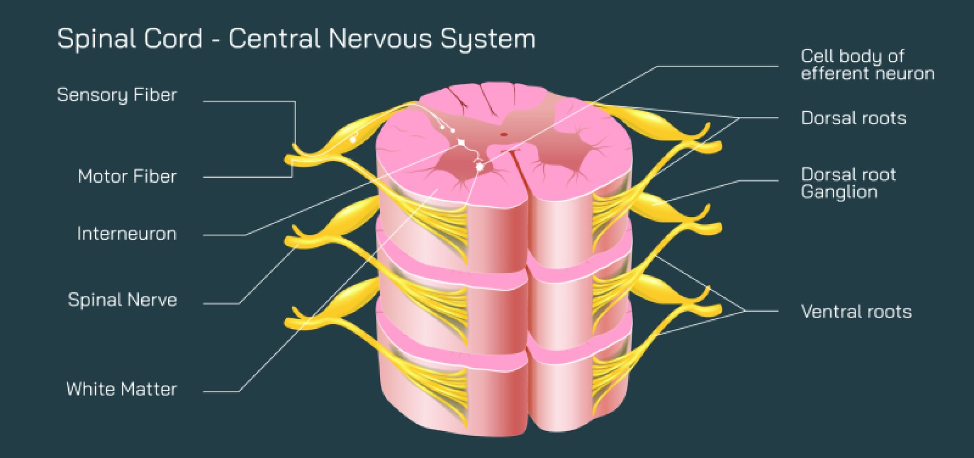
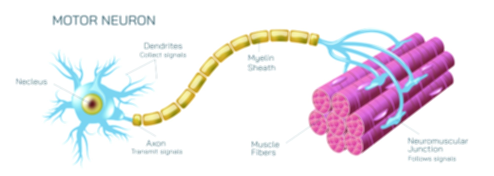
Motor neurons are specialized nerve cells responsible for transmitting signals from the brain or spinal cord to muscles and glands. These neurons enable voluntary movements, such as walking, writing, and speaking, as well as involuntary actions, like reflexes. Motor neurons connect with muscles at junctions called neuromuscular junctions, where electrical signals from the neuron prompt muscle fibers to contract.
Synapses are the communication points between neurons or between neurons and muscles. At a synapse, the electrical signal traveling along a neuron triggers the release of chemical messengers called neurotransmitters. These neurotransmitters cross a tiny gap known as the synaptic cleft and bind to receptors on the receiving cell, which can be another neuron or a muscle cell. This chemical communication is essential for signal transmission, coordination, and proper functioning of the nervous system.
Neurotransmitters are chemicals such as acetylcholine, dopamine, serotonin, and norepinephrine, each with specific roles in the body. Acetylcholine, for example, is critical for muscle contraction and motor neuron communication, while dopamine influences movement, motivation, and reward. Serotonin and norepinephrine play key roles in mood regulation and attention. The precise release, binding, and reuptake of neurotransmitters ensure smooth and accurate signal transmission across neural networks.
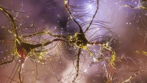
Visualizing these processes with 3D illustrations or vector diagrams helps students, researchers, and healthcare professionals understand the complexity of neural communication. Illustrations can show motor neurons extending from the spinal cord to muscles, synapses bridging neurons, and neurotransmitters crossing synaptic gaps. Such visualizations make abstract biological processes tangible and easier to comprehend, aiding learning and education.
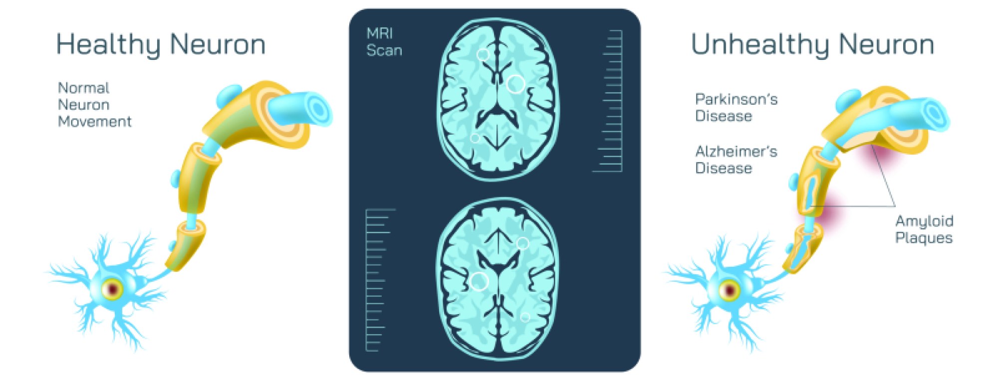
The study of neurotransmitters, motor neurons, and synapses is not only critical for understanding normal physiology but also for medical applications. Disruptions in these systems can lead to neurological disorders such as Parkinson’s disease, amyotrophic lateral sclerosis (ALS), myasthenia gravis, and depression. Illustrations of these components can assist in explaining disease mechanisms, therapies, and research interventions in neurology and neuroscience.
In conclusion, neurotransmitters, motor neurons, and synapses work together to transmit nerve signals from the brain and spinal cord to muscles and other target tissues. Through the coordinated action of electrical impulses, chemical messengers, and specialized junctions, the nervous system enables movement, communication, and bodily regulation. Illustrations of these systems enhance understanding, education, and awareness of how intricate neural networks control human and animal behavior and physiology.