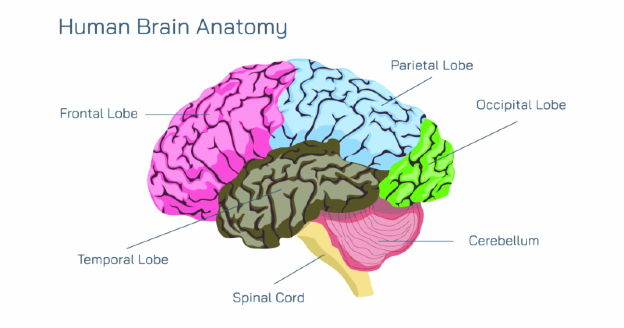
Brain Anatomy Showing Cerebrum, Cerebellum, Brainstem, Lobes, and Functional Regions of the Human Brain
The human brain is a complex biological control center that governs every physical, cognitive, emotional, and sensory function that defines life. Although protected within the skull and cushioned by membranes and cerebrospinal fluid, the brain contains distinct structural divisions that each serve specialized but interconnected roles. A detailed exploration of brain anatomy — including the cerebrum, cerebellum, brainstem, lobes, and key functional regions — reveals how individual neural systems blend together to generate thought, memory, movement, sensation, behavior, and identity. By examining the macrostructure of the brain alongside the rich internal communication networks that operate within it, the anatomy of this 1.3–1.4 kilogram organ becomes a map of human experience itself.
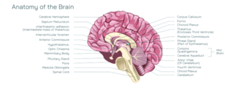
The largest portion of the brain is the cerebrum, making up more than 80 percent of total brain volume. It is divided into two hemispheres — left and right — linked by a thick bundle of neural fibers called the corpus callosum. This region is responsible for conscious thought, voluntary movement, language, reasoning, problem-solving, planning, judgment, emotions, and sensory interpretation. The cerebrum’s surface, the cerebral cortex, is deeply folded with gyri and sulci that increase surface area and allow a massive number of neurons to be packed into a relatively compact space. Beneath this surface lies white matter containing myelinated fibers that transmit signals rapidly between brain regions. Although the hemispheres look symmetrical, their functional specialization contributes to cognitive diversity: the left hemisphere is typically dominant for language, analytical reasoning, and logical processing, while the right hemisphere tends to excel in spatial awareness, emotional interpretation, creativity, and holistic thinking. Yet the hemispheres do not function in isolation — healthy cognition depends on continuous communication across both sides.
Within the cerebrum are four major lobes, each connected to specific functional capacities and shaped by evolution to handle distinct aspects of behavior and perception. The frontal lobe, positioned at the front of the brain, governs executive functions such as planning, decision-making, social behavior, impulse control, deliberate movement, and expressive language. It contains the prefrontal cortex — the most advanced region of human neural development — which manages long-term goals, moral reasoning, emotional regulation, and self-awareness. The parietal lobe, located behind the frontal region, integrates sensory information from the entire body and constructs the internal map of space, helping the brain understand touch, temperature, pressure, pain, and spatial orientation. The temporal lobe, found on the sides of the brain near the ears, houses neural circuits for hearing, language comprehension, auditory memory, emotional memory, and aspects of facial and object recognition. The occipital lobe, positioned at the back of the brain, is the visual processing hub responsible for interpreting shapes, color, depth, motion, light patterns, and spatial visual understanding. These lobes are not isolated modules — they interact constantly. For example, when a person views an object, the occipital lobe decodes the image, the temporal lobe identifies what it is, the parietal lobe determines its location in space, and the frontal lobe decides how to respond.
Directly beneath the cerebrum sits the cerebellum, a structure often associated primarily with balance but in truth handling an extensive array of functions related to precision. Although much smaller than the cerebrum, the cerebellum contains nearly half of all neurons in the brain, allowing it to coordinate and fine-tune movement with remarkable accuracy. It receives continuous input from sensory systems, the spinal cord, and other brain regions, comparing intended motion with executed motion and adjusting muscle activity and posture in real time. This makes the cerebellum crucial for smooth motor control, coordination, gait stability, and learned motor skills such as writing, playing instruments, and sports movements. It also plays an emerging role in cognitive timing, emotional regulation, motor learning, and aspects of automatic skill memory. In simple terms, the cerebrum forms the plan for movement while the cerebellum shapes that plan into fluid, efficient action.
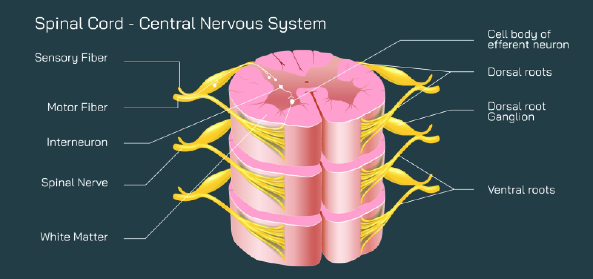
Connecting the brain to the spinal cord is the brainstem, an evolutionarily ancient structure that sustains the basic functions of life. Consisting of the midbrain, pons, and medulla oblongata, the brainstem controls involuntary vital operations such as breathing, heartbeat, blood pressure, swallowing, reflexes, body temperature, sleep-wake cycles, and the regulation of autonomic nervous system responses. It serves as the communication highway for all neural signals passing between the brain and the body. Damage to the brainstem can threaten survival not because it disrupts thinking but because it interrupts the biological rhythms that make consciousness possible in the first place. While the cerebrum governs the richness of experience, the brainstem ensures that physiological existence is maintained moment by moment.
Within and between these major regions are specialized functional networks that further refine the brain’s capabilities. The motor cortex, situated in the frontal lobe, sends signals to skeletal muscles to initiate voluntary movement. Just behind it, the somatosensory cortex in the parietal lobe processes sensation from skin, muscles, and joints, generating bodily awareness. The Broca’s area and Wernicke’s area, located primarily in the left hemisphere, manage speech production and language comprehension. The auditory cortex in the temporal lobe processes sound, while the visual cortex in the occipital lobe converts raw light signals into meaningful images. The limbic system, which includes the amygdala, hippocampus, and related structures, integrates emotion, memory formation, survival instincts, motivation, and reward processing. These functional regions do not define consciousness in isolation but form interdependent loops of perception, interpretation, memory, emotion, and action.
The brain’s overall function lies in integration. Sensory inputs arrive continuously from sight, sound, smell, taste, proprioception, and touch. The brain analyzes them, compares them to stored memories, weighs emotional relevance, predicts consequences, makes decisions, and initiates responses. This happens in fractions of a second, and the processing is distributed across networks rather than centralized in one location. A simple task, such as catching a falling object, involves coordination between visual centers identifying the object, parietal regions mapping its trajectory, motor regions planning the movement, the cerebellum adjusting muscle contractions, and the brainstem regulating cardiovascular and respiratory support. Every thought, every movement, and every emotional experience reflects this layered, synchronized activation spanning the cerebrum, cerebellum, and brainstem.
What makes the human brain especially remarkable is its ability to adapt and learn. Neural plasticity — the capacity to form new connections, strengthen frequently used pathways, and reorganize itself after injury — underlies the acquisition of new skills and the healing of damaged circuits. This adaptability operates from infancy through old age. During childhood and adolescence, the brain grows new connections rapidly in regions responsible for language, learning, and social behavior. In adulthood, neural wiring becomes more specialized and efficient as past experiences reinforce stable networks. Even in aging, the brain retains the ability to form new synaptic pathways when stimulated cognitively, socially, and physically.
The macro-anatomy of the brain — the cerebrum, cerebellum, brainstem, and lobes — provides the architecture. The functional regions within the lobes provide the specialization. Neural circuits, synapses, neurotransmitters, and electrical signaling provide the mechanism. Together, they produce memory, creativity, emotion, perception, intelligence, personality, reflex, instinct, and awareness. Without any single one of these structures the human experience would collapse: the cerebrum is required for thought and decision-making, the cerebellum for movement precision, the brainstem for life-sustaining functions, and the lobes for processing and integrating the world moment by moment.
The brain is therefore not just an organ of control — it is the biological seat of identity, regulating every action from heartbeat to imagination. Whether the task is reading a book, recalling a childhood memory, solving a problem, coordinating muscles during walking, feeling joy or fear, or simply breathing during sleep, different regions of the brain work together in continuous harmony. The anatomy of the brain reveals not only how life is maintained but how consciousness is built and how each person experiences reality uniquely through the coordinated work of their neural structures.
Through the structural and functional relationships among the cerebrum, cerebellum, brainstem, lobes, and specialized cortical regions, the human brain stands as the most sophisticated biological system known — one that translates microscopic electrical impulses into thought, action, memory, and meaning.