Spinal Cord and Central Nervous System: Structure, Function, and Neural Pathways Explained
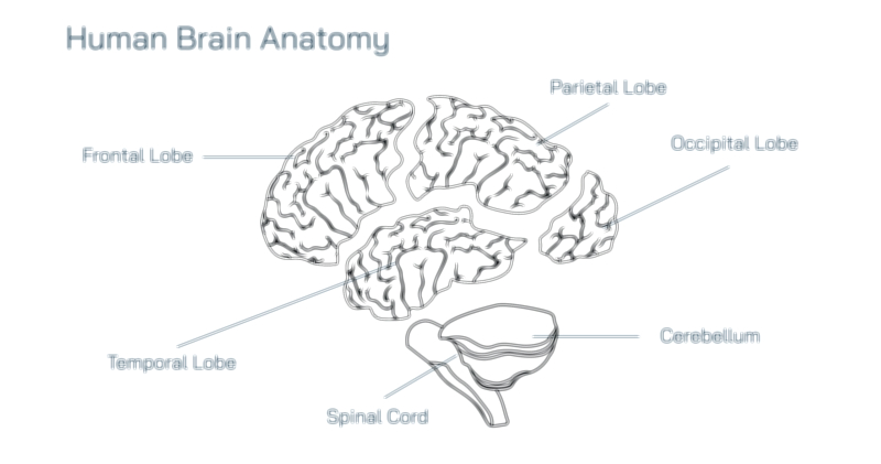
The spinal cord is one of the most vital and complex structures in the human body, serving as the primary communication bridge between the brain and the rest of the nervous system. Together with the brain, it forms the central nervous system (CNS), a sophisticated command center that governs every thought, movement, sensation, and autonomic function. While the brain performs perception, cognition, and higher decision-making, the spinal cord acts as the conduit through which motor instructions descend to the body and sensory information ascends to the brain. It also plays an independent role in reflexes, pain modulation, tissue coordination, and autonomic regulation, demonstrating that the spinal cord is not merely a passive cable but a dynamic neural processing center. It is remarkable that a structure roughly the diameter of a finger contains the wiring required to control millions of actions across a lifetime without interruption, even adjusting its function during injury, growth, and learning. Understanding the spinal cord and its relationship to the CNS requires exploring its anatomy, its internal pathways, and the mechanisms that allow the nervous system to function as a single integrated continuum.
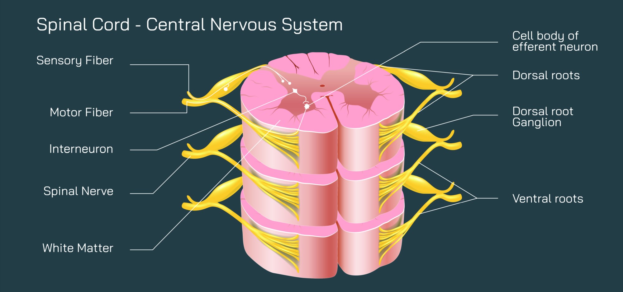
Anatomically, the spinal cord extends from the base of the brainstem through the protective canal formed by the vertebral column, typically reaching the level of the lower back before tapering into a bundle of nerve roots called the cauda equina. Although the spine itself continues downward, the spinal cord ends near the first or second lumbar vertebra, with long nerve roots traveling beyond this point to reach the lower body. At each segment along the spinal cord, right and left spinal nerves exit the vertebral column through small openings to supply specific regions of the body. These nerves are organized into cervical, thoracic, lumbar, sacral, and coccygeal segments, each corresponding to particular areas of motor and sensory control. Sensory nerves carry touch, temperature, pain, and proprioception back toward the spinal cord, while motor nerves transmit impulses from the CNS to muscles. This segmentation ensures that every part of the body has a dedicated neural link to the spinal cord, which feeds into the brain. The interdependence between spinal nerves and spinal cord segments becomes clear in clinical observation: damage to one region of the spinal cord predictably affects function in the corresponding part of the body below the injury.
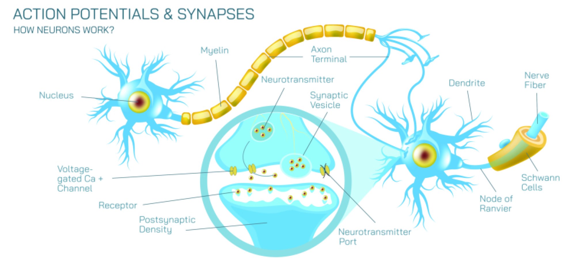
Inside the spinal cord lies a distinct structural arrangement of gray matter and white matter, forming a butterfly-shaped pattern that reflects how different types of neural information are processed. The inner gray matter contains neuronal cell bodies and synapses, where electrical signals are integrated and modified. The outer white matter consists primarily of myelinated axons arranged vertically into ascending and descending tracts, which act as highways for long-distance electrical transmission. Ascending tracts deliver sensory information to the brain, while descending tracts carry motor commands from the cerebral cortex and brainstem to the body. This dual traffic system makes the spinal cord both a communication bridge and a computational hub. The organization of gray matter into dorsal (posterior) horns and ventral (anterior) horns is functionally important: dorsal horns receive incoming sensory information, while ventral horns house the motor neurons that directly activate muscle contraction. Between these regions lies a complex network of interneurons that regulate reflexes and modulate signals. At the structural level alone, the spinal cord demonstrates how anatomy supports fast communication while also enabling local control.
The spinal cord’s neural pathways work through carefully organized circuits that maintain the CNS’s functional unity. The ascending sensory pathways include major systems such as the dorsal column-medial lemniscus pathway, which carries fine touch and proprioception, and the spinothalamic pathway, which carries pain and temperature signals. These pathways ensure that sensory information from the body reaches the brain with extraordinary detail and spatial accuracy. They allow the brain to determine where the body is in space, the texture of objects, thermal conditions, and the detection of harm. At the same time, the descending motor pathways — especially the corticospinal and reticulospinal tracts — transport motor instructions from the brain to the spinal cord. These pathways enable voluntary movement, muscle coordination, and postural control. Even simple actions such as reaching for a cup require rapid integration of brain signals with spinal feedback to adjust muscle force and trajectory in real time. The spinal cord accomplishes these computations continuously and automatically, ensuring that motion appears fluid and effortless.
One of the most striking capabilities of the spinal cord is its ability to perform reflex actions independently of the brain. A reflex arc involves sensory receptors detecting a stimulus and immediately activating motor neurons without requiring conscious processing. For example, touching a hot surface results in an instantaneous withdrawal of the hand before pain is even perceived in the brain. This rapid response is vital for protection from injury and demonstrates that the spinal cord contains built-in neural circuits capable of initiating action. Reflexes are not isolated from brain control — the brain can modulate reflex intensity through descending pathways — but they reveal the dual nature of the spinal cord as both an autonomous processor and a channel for higher commands. Reflex circuits also contribute to posture, locomotion, and muscle tone, coordinating complex activities such as walking through rhythmic activation patterns in spinal locomotor networks.
Beyond sensory and motor roles, the spinal cord also supports autonomic functions, regulating internal organs through sympathetic and parasympathetic pathways. Nerve fibers in the spinal cord influence heart rate, blood vessel diameter, sweating, digestive processes, and bladder control. These autonomic signals allow the CNS to maintain physiological stability without conscious awareness. Emotional states and stress also affect spinal autonomic output, demonstrating that the CNS coordinates bodily function holistically rather than in isolated segments. Conditions such as spinal cord injury help illustrate how deeply the spinal cord contributes to autonomic life processes: damage can interrupt signals regulating blood pressure, body temperature, and organ function, showing that the spinal cord is just as vital to involuntary regulation as it is to voluntary movement.
The relationship between the spinal cord and the brain is reinforced by their shared protective environment — the meninges and cerebrospinal fluid (CSF) — which cushion the CNS from physical impact and help maintain a stable chemical environment. The dura mater, arachnoid mater, and pia mater form three tough membranes encasing the spinal cord, while CSF circulates through the subarachnoid space to absorb shocks and deliver nutrients. The spinal cord is additionally protected by the bony vertebrae that surround it, though this structural confinement makes the neural tissue highly vulnerable to compression or displacement. Even minor swelling or narrowing of the spinal canal can compress the cord or its nerves, producing sensory loss, weakness, or pain. The delicate partnership between flexibility, protection, and neural conduction is continuously maintained through spinal biomechanics and fluid dynamics.
The spinal cord’s developmental and adaptive characteristics also contribute to its function. During embryonic growth, the spinal cord establishes neural circuits before birth and later adapts through plasticity — the ability to strengthen, weaken, or reorganize pathways based on experience. This plasticity is essential for learning motor skills, recovering from injury, and adapting reflexes and coordination across the lifespan. Rehabilitation after spinal injuries relies largely on encouraging remaining neural pathways to reorganize and take over lost functions through repetitive training and sensory stimulation. Although the CNS cannot regenerate neurons as extensively as peripheral nerves, it retains the capacity to remodel synapses, modify circuits, and optimize function when given the appropriate biological environment.
The importance of the spinal cord becomes especially clear when injury or disease disrupts its functioning. Trauma, degenerative disc disease, tumors, infections, autoimmune disorders, and congenital malformations can damage neural pathways. Because the spinal cord organizes function from top to bottom, the level of injury determines the extent of dysfunction. Damage high in the cervical cord affects the arms, legs, diaphragm, and autonomic control, whereas injuries lower in the thoracic or lumbar cord may affect only the legs or organs below the injury level. Still, the full spectrum of spinal cord disorders illustrates how many systems rely on the continuity of CNS pathways: sensation, movement, breathing, digestion, blood pressure, bladder and bowel control, and even thermoregulation depend on spinal integrity. Advances in neurology, robotics, exoskeleton technology, neural implants, and stem-cell therapies continue to expand possibilities for restoring function, reflecting scientific determination to heal an organ that plays such a defining role in life.
Ultimately, the spinal cord stands as a testament to the elegance and efficiency of biological design. It serves as a high-speed communication highway, a reflex generator, a locomotor coordinator, an autonomic regulator, and a vital extension of the brain. Through its ascending and descending pathways, it links every muscle and sensory receptor to the central command system, enabling movement, perception, and homeostasis. Together with the brain, it forms the central nervous system — the network that allows humans to interact with, interpret, and respond to the world. Without the spinal cord’s seamless integration of structure and function, thought could not become action, sensation could not become experience, and life in its dynamic, responsive form would not exist.