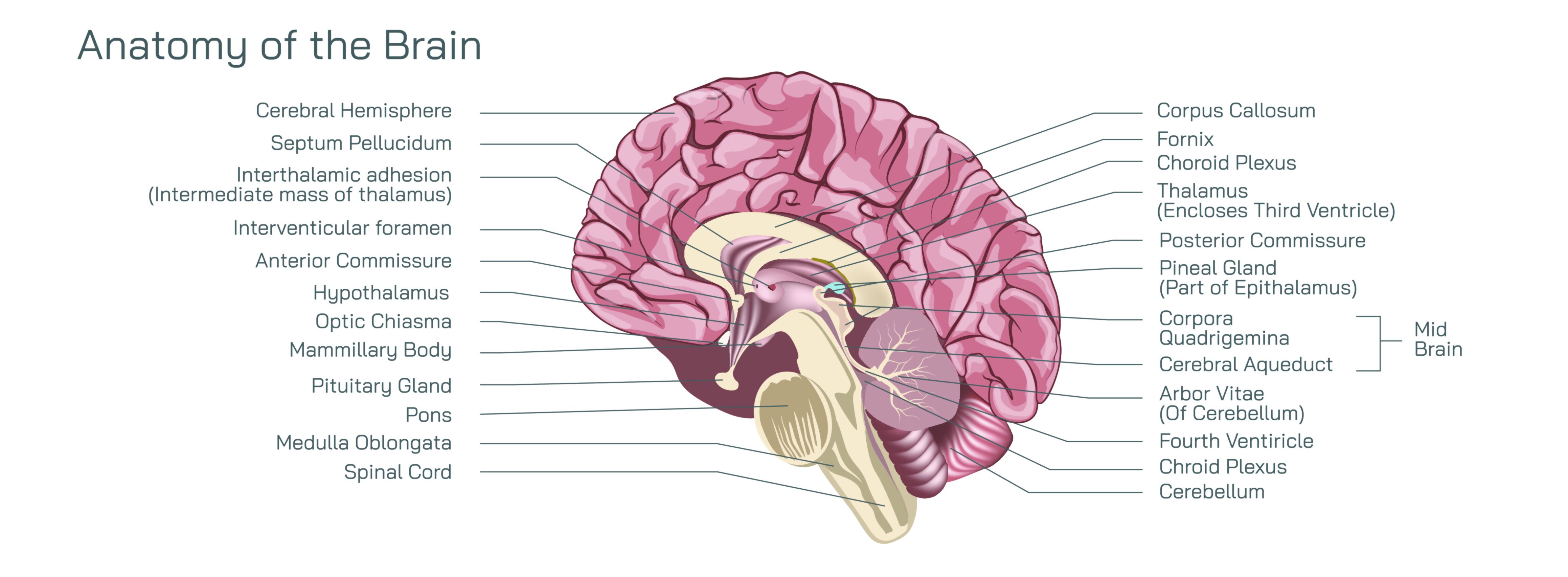
Anatomy of the Brain Showing Major Lobes, Neural Pathways, and Functional Regions of the Human Nervous System
The human brain is the most intricate and powerful organ known in biology, orchestrating every sensation, emotion, thought, movement, and memory that defines human experience. Its structure reflects millions of years of evolutionary refinement, shaping a network capable of perception, creativity, abstract reasoning, social connection, physical coordination, and survival. When viewed anatomically, the brain is not a single uniform mass but a complex landscape divided into specialized lobes, interconnected by dense neural pathways, and governed by functional regions that communicate continuously. The brain’s external surface displays a folded pattern of gyri and sulci that dramatically increase cortical surface area and cognitive capacity, while its internal fiber networks form the highways of the nervous system, enabling trillions of signals to travel every second. The study of brain anatomy reveals not only where structures exist, but how they cooperate to generate consciousness, behavior, and personality.
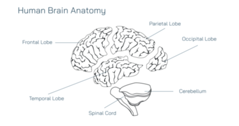
Four major lobes dominate the cerebral cortex, each sitting in its own territory and contributing to different categories of brain function. At the forefront and occupying the largest area of the cortex is the frontal lobe, widely recognized for its role in executive control, decision-making, speech, planning, logic, motivation, emotional regulation, and voluntary movement. It houses the prefrontal cortex, the seat of personality and social behavior, where judgment and long-term goal planning are shaped by learned experience and moral reasoning. The frontal lobe also contains the primary motor cortex, a strip of tissue responsible for sending motor commands to muscles throughout the body. This region organizes movement in a somatotopic pattern—meaning specific territories of the cortex correspond to specific body parts. On the left hemisphere of most individuals lies Broca’s area, essential for speech production and verbal articulation. Injury to the frontal lobe often leads to drastic changes in behavior, emotional control, and impulse management, revealing its essential role in shaping identity and rational thought.
Immediately behind the frontal lobe rests the parietal lobe, the perceptual and integrative region where sensory input from the body becomes meaningfully interpreted. The primary somatosensory cortex receives tactile information such as touch, temperature, texture, pressure, and pain, mapping the entire body in a spatial grid that mirrors the motor cortex. Beyond basic sensation, the parietal lobe supports spatial awareness, navigation, and proprioception — the internal sense of limb position and body orientation. It also integrates visual information with movement, enabling coordinated actions such as reaching, grasping, or walking around obstacles. Higher parietal regions contribute to mathematical reasoning, symbolic processing, and logical organization. When this lobe is damaged, individuals may struggle to recognize their own limbs, navigate familiar environments, or understand spatial relationships, demonstrating that perception is far more than raw sensory input; it requires cortical interpretation.
Toward the back of the brain lies the occipital lobe, the primary visual center of the nervous system. Although sight begins in the eyes, true visual experience is constructed in the occipital cortex, where light signals received from the retinas are processed into edges, colors, depth, motion, and symbolic meaning. The primary visual cortex (V1) performs the first step in decoding images, while surrounding areas identify patterns, faces, shapes, letters, and movement. These visual signals then travel forward to regions that integrate them with memory, emotion, and decision-making. Because the neural pathways crossing between the eyes and brain are highly organized, injury to a specific stripe of occipital cortex can cause blindness in a corresponding area of the visual field. This lobe is the perfect example of how sensation requires cortical interpretation — without the occipital cortex, sight ceases even if the eyes remain intact.
On the lateral side, beneath the temples, lies the temporal lobe, responsible for auditory processing, language comprehension, long-term memory formation, and emotional learning. The primary auditory cortex interprets sound patterns such as speech, music, and environmental noise, while Wernicke’s area — usually on the left — allows language to be understood, linking words to meaning. The temporal lobe also holds the hippocampus deep within its medial region, forming the structural basis of memory storage and recall. Experiences passing through the senses become encoded into memories through hippocampal circuitry, while emotion-laden memories are strengthened through connections to the amygdala. Thus the temporal lobe forms a crossroads between perception, memory, and emotional response. Damage here may result in memory loss, difficulty recognizing faces or sounds, disturbances in language comprehension, or emotional volatility.
Although the four lobes of the cerebral cortex provide the most visible structure of the brain, their function depends on the seamless interplay of neural pathways that transfer information between them. These pathways are composed of white matter tracts — bundles of myelinated axons that act like electrical wiring. Among the most important pathways is the corpus callosum, which bridges the left and right hemispheres, allowing both sides of the brain to cooperate constantly. Without this connection, each hemisphere would operate independently, impairing coordination between creative and logical processes. Other notable pathways include the arcuate fasciculus, a fiber tract connecting Broca’s area and Wernicke’s area, enabling speech production and language comprehension to operate as a unified system. The optic radiations deliver visual information from the thalamus to the occipital lobe, while the cingulum bundle links limbic regions to the frontal cortex, making emotional context available to conscious decision-making. These neural highways form the backbone of cognition, symbolizing the brain as a network rather than a collection of isolated parts.
Beneath the outer lobes sits the subcortical functional brain, home to deeper regions that coordinate emotion, movement, memory, and survival responses. The thalamus, often called the gateway to the cortex, acts as a sensory relay station, directing incoming information from the eyes, ears, skin, and internal organs to the appropriate cortical regions. The basal ganglia, a cluster of interconnected nuclei, regulate voluntary movement, procedural learning, and reward-driven behavior. When functioning properly, they produce smooth coordinated motion; when impaired — as in Parkinson’s disease or Huntington’s disorder — movement becomes rigid, tremulous, or uncontrolled. The cerebellum, positioned below the occipital lobe, further refines movement by calibrating balance, timing, and precision. Even though the cerebellum is not part of the cortex proper, its contribution to fluid motor control is so vital that damage can result in unsteady posture, incoordination, or slurred motion patterns.
The brainstem — consisting of the midbrain, pons, and medulla oblongata — anchors the nervous system to essential life functions. It regulates heartbeat, breathing, digestive reflexes, sleep-wake cycles, and automatic responses. While the cortex gives rise to higher thought and identity, the brainstem ensures survival by maintaining vital physiological rhythms from birth to death. The integration of the cortex, limbic system, basal ganglia, cerebellum, and brainstem reflects the harmonious architecture of the nervous system: high-level cognition, emotional learning, instinct, and bodily regulation coexist through constant neural communication.
A fully labeled diagram of brain anatomy therefore illustrates much more than the borders of lobes; it depicts a hierarchy of interacting systems. The frontal lobe shapes behavior and decision-making; the parietal lobe decodes the world through sensation and spatial orientation; the occipital lobe transforms light into vision; the temporal lobe translates sound into meaning and memory. Neural pathways unite these lobes into a seamless cognitive organism where sensory signals become perceptions, perceptions become experiences, experiences become memories, and memories inform future decisions. Subcortical regions sustain emotion, motivation, and motor control, while the brainstem preserves the fundamental rhythms of life.
To study brain anatomy is to bridge structure and function — to understand how matter generates mind. The human brain is not simply an organ; it is the biological foundation of personality, learning, autonomy, imagination, and connection. Every lobe, every pathway, and every functional region contributes to the orchestration of consciousness, creating a system capable of thought, movement, empathy, planning, creativity, and resilience. The anatomy of the brain reveals the architecture of human existence, capturing in physical form the processes that make every experience and every identity uniquely alive.