Peripheral Vestibular System Anatomy: Balance and Inner Ear Function Explained
The peripheral vestibular system forms the sensory foundation for balance, spatial orientation, and stabilization of vision during movement, and understanding it begins with a close look at the anatomy of the inner ear. Although balance might feel effortless in daily life, it relies on a highly specialized network of receptors within the temporal bone that constantly detect motion, head position, and gravitational forces, sending signals to the brain so posture and eye movements can be precisely adjusted in real time. A comparison between the structural layout and functional roles of the semicircular canals, otolith organs, vestibular nerve, and supporting structures demonstrates how the peripheral vestibular system converts the mechanical language of motion into the electrical signals that guide equilibrium.
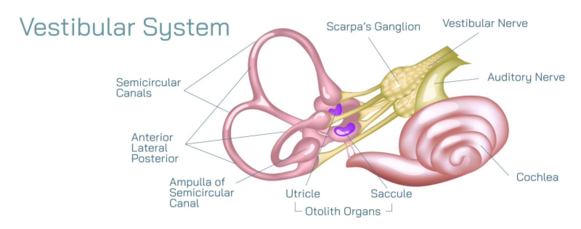
At the core of the vestibular system is the bony labyrinth, a complex series of hollow chambers and looping canals embedded deep in the skull. Within this rigid outer casing lies the membranous labyrinth, a fluid-filled network suspended inside the bony labyrinth. The spaces between the two are filled with perilymph, while the fluid inside the membranous labyrinth is called endolymph. This separation of fluid environments is essential because changes in the movement of endolymph inside the membranous structures serve as the primary physical input for detecting motion. The vestibular organs that sense head movement and gravitational pull are divided into two categories: the semicircular canals, which detect rotational acceleration, and the otolith organs, which sense linear acceleration and static head position relative to gravity.
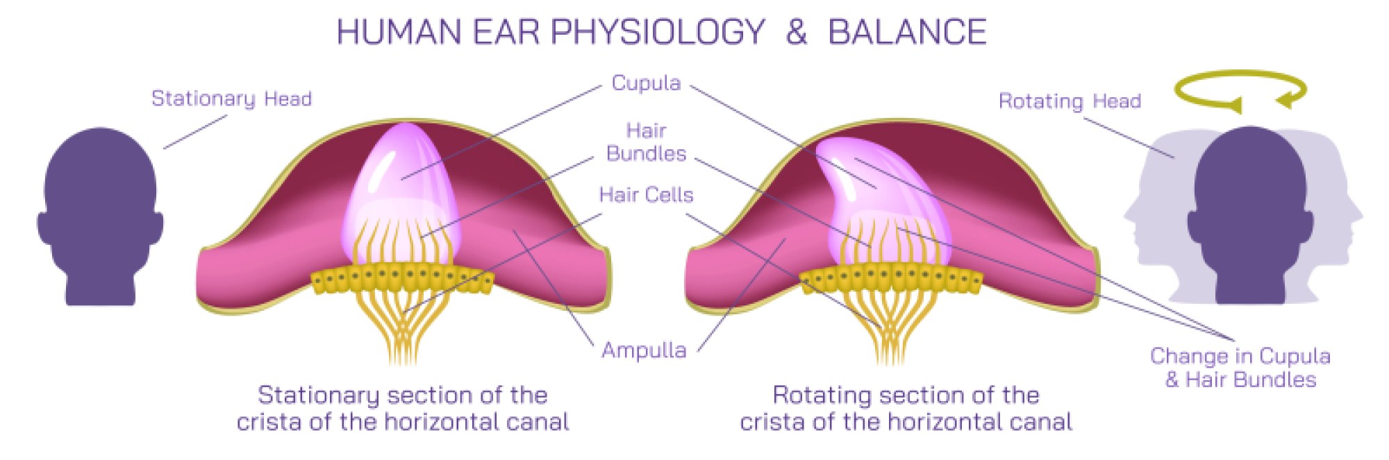
The three semicircular canals — anterior (or superior), posterior, and horizontal — are arranged almost at right angles to one another, allowing the detection of rotation in three dimensions. Each canal forms a loop connected to the vestibule and houses an expanded region called the ampulla. Within each ampulla lies the crista ampullaris, a raised structure containing hair cells, the sensory receptors of the vestibular system. These hair cells are topped with fine, hair-like projections called stereocilia that extend into a flexible gelatinous structure known as the cupula. When the head rotates, inertia causes the endolymph inside the canal to lag slightly behind the motion of the canal walls, bending the cupula and deflecting the stereocilia. This mechanical bending alters ion channel permeability in the hair cells, converting physical motion into neural signals. The brain interprets increased or decreased firing rates along branches of the vestibular nerve to determine the direction and speed of rotation.
While the semicircular canals detect angular motion, the otolith organs — the utricle and saccule — specialize in sensing linear acceleration and gravity. These organs contain sensory patches known as maculae, which resemble the cristae of the canals in their use of hair cells but differ in structure and functional triggers. Overlying each macula is a gelatinous layer embedded with tiny calcium carbonate crystals known as otoconia. The weight of these crystals gives the gelatinous sheet greater inertia. When the head tilts or the body accelerates linearly, gravity or momentum causes the otolithic membrane to shift relative to the hair cells beneath it. This displacement bends the stereocilia, producing neural signals that convey changes in head orientation or straight-line motion. Because the utricle is oriented horizontally and the saccule vertically, the pair can detect motion in any linear direction, including up-and-down, side-to-side, and forward-and-back.
The information gathered from the semicircular canals and otolith organs converges into the vestibular nerve, one of the two divisions of the eighth cranial nerve. From the vestibular nuclei in the brainstem, the signals are integrated with visual and proprioceptive information from the eyes and body. This integration allows the brain to instantly recalibrate posture, coordinate limb positioning, and maintain stability even during complex or abrupt movement. One of the most spectacular outcomes of vestibular processing is the vestibulo-ocular reflex, an automatic mechanism that keeps visual images stable on the retina even while the head moves. Through this reflex, the vestibular system drives rapid compensatory eye movements in the opposite direction of head rotation, allowing us to read, walk, or look around without the world appearing to blur.
The everyday actions of standing upright, turning the head, bending over, or shifting direction while walking all depend not only on the vestibular apparatus itself but also on the continual comparison of vestibular, visual, and proprioceptive inputs. When these systems agree with each other, balance feels smooth and automatic. When they conflict, the brain struggles to determine which information to trust. This mismatch explains why motion sickness and vertigo occur when the vestibular organs signal motion while the eyes report stability, such as during travel, virtual simulation, or inner ear disturbances. In pathological states like vestibular neuritis, Ménière’s disease, or benign paroxysmal positional vertigo, disruptions within the peripheral vestibular system interfere with fluid movement or sensory receptor functioning, leading to dizziness, imbalance, nausea, visual instability, and difficulty walking.
Despite its small size, the peripheral vestibular system must work with exceptional precision, because even a tiny misinterpretation of head movement can cause sway, staggering, or visual disturbance. The semicircular canals detect rotation, the otolith organs monitor linear motion and gravity, the hair cells transduce mechanical bending into electrical impulses, and the vestibular nerve delivers those signals to the brain. Together, these structures encode the full three-dimensional dynamics of body movement. When the anatomy of the vestibular system is viewed through diagrams that illustrate the arrangement of the canals, otolithic organs, and neural pathways, it becomes clear that balance is not an incidental or secondary ability — it is the product of highly evolved sensory engineering. The peripheral vestibular system forms the silent stabilizer that allows every human action, from standing still to running at full speed, to feel natural, coordinated, and visually anchored in the world.