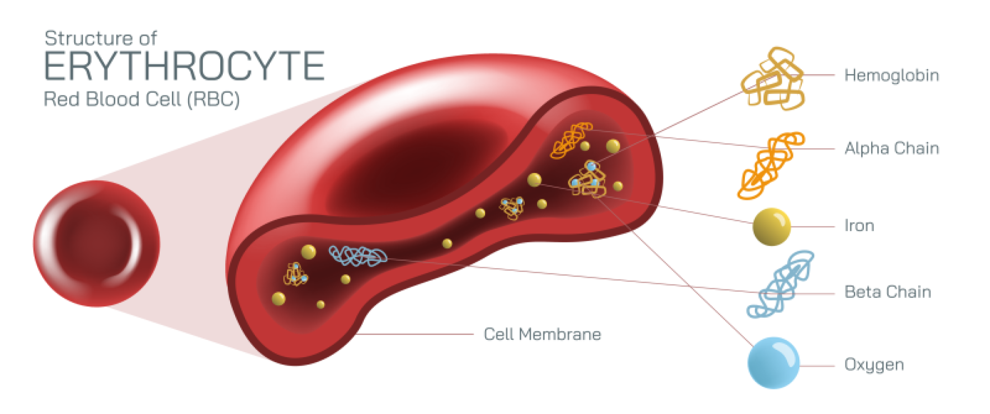
Antigen vs Antibody Illustration Showing Immune System Interaction and Defense Mechanism
An antigen vs antibody illustration serves as a powerful way to visualize the protective strategy of the immune system, translating a complex biochemical defense into a clear picture of recognition and response. At the most fundamental level, an antigen is any foreign substance—such as a virus particle, bacterium, parasite, toxin, allergen, or even an abnormal cell—that carries molecular structures recognized by the immune system as “non-self.” These molecular structures are often proteins or carbohydrate patterns located on the surface of the invading particle. In contrast, an antibody is a Y-shaped protein produced by specific immune cells (B lymphocytes) that circulates through the bloodstream and tissues to neutralize or flag antigens for destruction. Observing these two elements side by side reveals a dynamic relationship rather than simply two separate biological entities: one triggers the immune alarm, the other responds with precision targeting.
A realistic illustration of the interaction typically begins with the antigen. In a diagram, an antigen appears as a distinct foreign particle—perhaps a virus with spikes on its surface, or a bacterial cell studded with proteins and sugar chains. The key visual detail lies in the epitopes, which are specific molecular shapes or “binding sites” located on the antigen’s surface. These epitopes act like molecular fingerprints that distinguish one pathogen from another. The immune system reads these shapes through antibody receptors, and the illustration frequently highlights one epitope being recognized by the matching tip of an antibody. This matching relationship forms the core of antigen–antibody interaction: each antibody is uniquely shaped to bind to a specific epitope, fitting into place much like a lock and key. In diagrams, this connection is shown by the forked ends of the antibody—the antigen-binding sites—aligning perfectly with the epitope’s contour.
On the other side of the illustration, the antibody takes center stage. It is typically drawn in the iconic Y-shape, with two identical arms (Fab regions) that contain antigen-binding sites and a stem (Fc region) that connects the antibody to the broader immune system. The arms bind the antigen, while the stem signals immune cells that a threat has been captured. This dual function is depicted in many diagrams by showing immune cells—such as macrophages or natural killer cells—interacting with the Fc region once the antigen has been bound. This visual emphasis communicates the full purpose of antibodies: they not only identify intruders but also mobilize the immune attack.
When antigen and antibody are shown together in an illustration, the steps of the immune response become clearer:
Recognition – The antibody finds and binds to the antigen’s epitope with precise molecular compatibility.
Neutralization – By binding to the antigen, the antibody may block the pathogen’s ability to infect host cells. For example, antibodies covering the spikes of a virus prevent it from docking onto cell receptors.
Agglutination (clumping) – Antibodies can bind multiple antigens simultaneously, linking them into groups that are easier for immune cells to eliminate.
Opsonization and signaling – The Fc region of the antibody triggers immune cells such as macrophages to engulf and destroy the antibody-tagged pathogen.
Complement activation – Bound antibodies may also activate a cascade of blood proteins that directly destroy pathogens.
These steps are often illustrated with arrows or sequential visual panels, allowing the viewer to see how a single binding event expands into a coordinated immune response. The antibody transforms from a passive molecule into an active communicator that unlocks powerful cellular and biochemical defenses.
The antigen vs antibody illustration also highlights how memory and immunity develop. Once the immune system encounters a particular antigen, memory B cells are formed that retain the blueprint for producing the same antibody in the future. In a diagram, these memory cells might be placed in a separate panel as long-term defenders waiting for reinfection. The next time the same antigen appears, the immune system can respond much faster, producing a rapid wave of antibodies that neutralize the threat before illness takes hold. This visual detail reinforces how vaccination works: vaccines introduce harmless antigens or fragments of pathogens to train the immune system to create antibodies and memory cells before real exposure occurs.
Another nuance often captured in illustrations is the difference between self and non-self recognition. Healthy human cells also display surface molecules, but the immune system has learned not to attack them. Only unfamiliar or abnormal molecules trigger antibody generation. When this discrimination fails, autoimmune diseases arise—conditions where antibodies target the body’s own tissue. Some textbooks or medical diagrams include an inset comparing normal immune recognition and autoimmune misrecognition to reinforce this principle of immune tolerance.
The importance of antigen–antibody interaction extends beyond infection and immunity; it plays a role in allergies, transplant compatibility, blood type matching, immunotherapy, and medical diagnostics. In allergies, for example, harmless substances like pollen or peanuts act as antigens that trigger antibody overreaction. In blood transfusions, mismatched antigens on red blood cells can bind to incompatible antibodies, causing dangerous clumping. In modern medicine, laboratory tests such as rapid diagnostic kits work by mimicking antigen–antibody binding and producing a visible signal when a match occurs.
Ultimately, an illustration comparing antigens and antibodies does more than label two biological components—it captures the choreography of immune protection. The antigen initiates the conflict, the antibody identifies and binds the threat, and the immune system mobilizes to eliminate danger. The precision of that binding interaction is the foundation of immune defense, ensuring that the body can distinguish friend from foe at the microscopic level. Through a simple image of alignment between a pathogen’s epitope and an antibody’s binding site, the viewer gains insight into how the immune system defends life using recognition, adaptation, and memory.