Motor Neuron Vector Illustration Showing Neuron Structure, Axon, Dendrites, and Impulse Transmission in the Nervous System
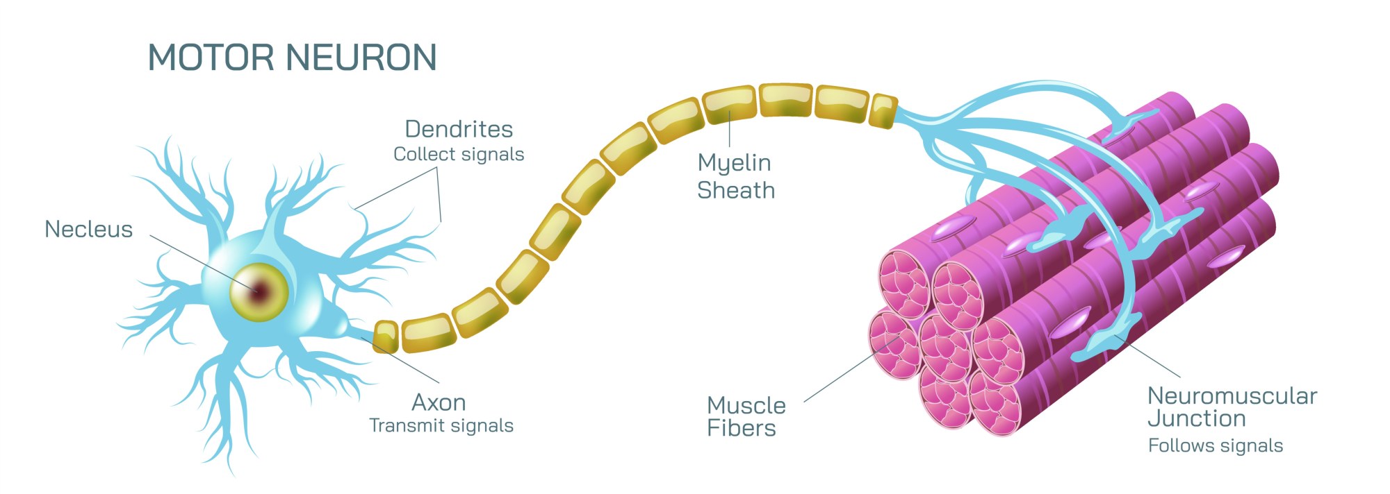
Motor neurons are specialized nerve cells responsible for transmitting electrical impulses from the central nervous system (CNS) to muscles and glands, facilitating voluntary and reflexive movements. Understanding their structure and function is fundamental in neurobiology, physiology, and medical education. A vector illustration of a motor neuron typically integrates cell body anatomy, dendritic input regions, axonal pathways, and synaptic terminals, providing a comprehensive and visually intuitive representation of impulse transmission. By combining labeled components, directional arrows, and magnified insets, such illustrations allow learners to understand both the cellular structure and functional mechanisms of motor neurons within the nervous system.
At the center of the illustration is the motor neuron cell body (soma). The soma is depicted with a spherical or oval shape, containing the nucleus, nucleolus, and cytoplasm. Labels identify each organelle, emphasizing their roles in protein synthesis, metabolic activity, and maintenance of neuronal function. Color coding differentiates the nucleus and cytoplasm from surrounding structures, enhancing clarity. Arrows indicate the integration of incoming signals from dendrites toward the soma, visually demonstrating the directional flow of electrical impulses.
Dendrites extend from the cell body, branching extensively to form a receptive network. Labels highlight these structures as primary input regions, receiving signals from other neurons via synapses. Arrows indicate the direction of electrical impulses toward the soma, emphasizing signal convergence. Vector insets may magnify synaptic junctions, showing neurotransmitter release from presynaptic neurons and receptor binding on dendritic membranes, visually linking microscopic synaptic events to macroscopic neuronal function. Color-coded gradients may indicate signal strength or intensity, illustrating how multiple inputs integrate to trigger action potentials.
The axon is illustrated as a long, singular projection extending from the soma, transmitting impulses away from the cell body toward effector muscles or glands. Labels identify the axon hillock, myelin sheath segments, nodes of Ranvier, and axon terminals. Arrows along the axon depict the propagation of action potentials, highlighting the unidirectional flow of electrical signals. Color gradients or animated markers may show impulse velocity, with insets explaining the role of myelination in saltatory conduction and efficient signal transmission.
Axon terminals or synaptic boutons are depicted at the distal ends of the axon, forming connections with muscle fibers or other target cells. Labels indicate neurotransmitter vesicles and synaptic clefts, emphasizing the role of chemical signaling in translating electrical impulses into muscular contraction or glandular secretion. Arrows show the release of neurotransmitters (e.g., acetylcholine) into the synaptic cleft, binding to receptors on the effector cell membrane, and initiating a physiological response. Magnified insets may illustrate vesicle fusion and neurotransmitter diffusion, linking cellular structure to functional output.
The vector illustration often includes functional pathways, such as signals originating from the CNS traveling through motor neuron axons to target muscles. Arrows indicate the flow of impulses from dendrites to soma, along the axon, and to synaptic terminals, visually reinforcing the concept of directional signal transmission. Labels may highlight key processes like action potential initiation, propagation, and synaptic transmission, providing a holistic view of neuron functionality.
Additional features may include comparative panels showing myelinated versus unmyelinated axons, illustrating differences in impulse conduction speed. Color-coded overlays may differentiate fast and slow pathways, and insets may show cross-sections of axons with nodes of Ranvier to explain saltatory conduction. Arrows may indicate how demyelination, as seen in certain neurological disorders, slows or blocks impulse transmission, connecting structure to clinical relevance.
By combining soma, dendrites, axon, axon terminals, synapses, and directional arrows for impulse flow, a motor neuron vector illustration provides a detailed understanding of neuronal anatomy and function. Color coding, labeled organelles, magnified synaptic insets, and functional pathways allow learners to visualize the connection between cellular structure and physiological response, linking microscopic anatomy to macroscopic nervous system activity.
Ultimately, a motor neuron vector illustration demonstrates the integration of structure and function in the nervous system, showing how electrical impulses are received, transmitted, and converted into action at effector sites. Through labeled dendrites, soma, axon, and synaptic terminals, along with directional flow indicators and magnified synaptic mechanisms, the diagram transforms abstract neurophysiological concepts into an educational, visually engaging, and intuitive tool for neuroscience learning, medical education, and physiological study.