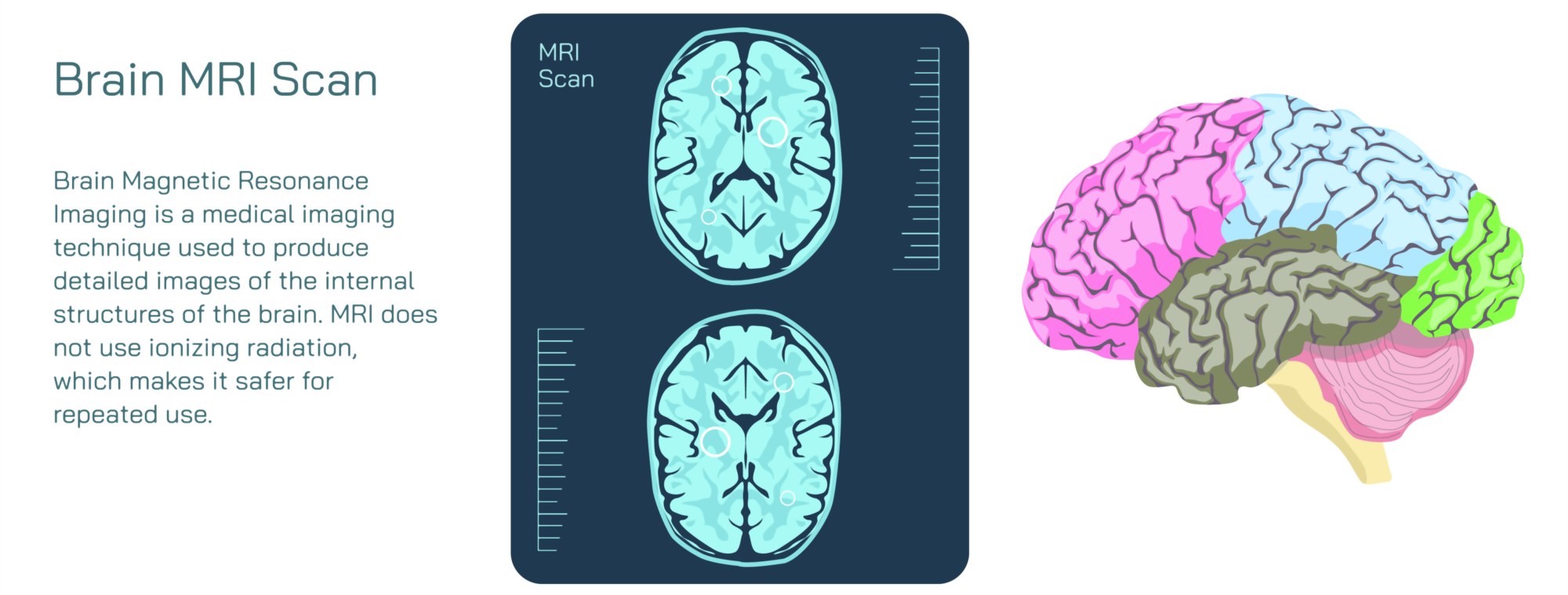
Brain MRI Scan Illustration: Cross-Section Imaging Showing Brain Structures and Pathologies
A brain MRI scan illustration serves as one of the clearest and most informative windows into the inner workings of the human nervous system, transforming what is normally hidden behind the skull into a detailed visual display of tissues, structures, and potential abnormalities. MRI—Magnetic Resonance Imaging—creates cross-sectional slices of the brain without requiring invasive procedures, and medical illustrations often emulate this appearance by showing layered images that look like thin sections from the top of the head to the base. These slices reveal gray matter, white matter, fluid-filled ventricles, blood vessels, and midline structures with a clarity that has revolutionized neurology and radiology. In a typical cross-section illustration, darker and lighter shades show how different tissues interact, and each major region stands out based on its unique appearance and contrast signal. The cerebral cortex forms a rippled outer layer, the white matter pathways appear as lighter internal bundles, and deeper structures such as the thalamus and basal ganglia emerge near the center of the image. The brainstem becomes visible toward the lower slices, connecting the brain to the spinal cord like a biological highway. The cerebellum, positioned at the back of the lower brain, stands out with its tightly folded surface designed for motor coordination and balance. A well-designed MRI illustration doesn’t simply mimic the look of the scan, but carries labels and shading that guide the viewer through this sophisticated anatomy with intuitive clarity.
When interpreted visually, MRI slices reflect the brain’s layered organization and reinforce how each region contributes to thought, movement, memory, and survival. In axial (horizontal) slices, the frontal lobes appear at the front of the scan, responsible for decision-making, emotion control, planning, and personality. The parietal lobes mark the upper middle portion of the cross-section, acting as centers for sensation, body awareness, and spatial mapping. The temporal lobes appear on either side of the scan, associated with language, hearing, and memory formation. The occipital lobe occupies the back, processing visual information and shaping perception. A sagittal slice, which cuts the brain into left and right halves, highlights the corpus callosum, the thick bridge of white matter connecting the two hemispheres and enabling constant information exchange. Meanwhile, a coronal slice—like looking at the brain from the front—reveals symmetry between left and right sides, while drawing attention to structures such as the amygdala and hippocampus deep within the medial temporal lobe. These structures regulate fear, emotional processing, and memory storage—core functions that directly influence human behavior and experience. Thus, each MRI cross-section teaches not only structural anatomy, but the functional architecture of human consciousness.
One of the core strengths of MRI illustration lies in its ability to distinguish between normal and abnormal patterns, which is why medical diagrams frequently include side-by-side comparisons of healthy and pathological scans. A healthy brain typically displays symmetry between left and right hemispheres, smooth cortical boundaries, balanced ventricle size, and uniform white-matter appearance. When pathology is present, these visual patterns change in identifiable ways. For example, a stroke often appears as a region of altered contrast—either hyperintense (bright) or hypointense (dark) depending on timing and MRI type—indicating tissue damage from disrupted blood supply. The location of the stroke correlates directly with neurological symptoms; a lesion in the left frontal lobe may cause speech impairment, whereas damage to the right parietal region may trigger neglect of the opposite side of space. A tumor on an MRI illustration might appear as an irregular mass that pushes or infiltrates neighboring tissue, distorts the ventricles, or disrupts symmetry. The surrounding brain may show swelling, depicted as areas of increased brightness and effaced sulci. In neurodegenerative diseases such as Alzheimer’s, the ventricles appear enlarged and cortical thickness diminishes as brain tissue gradually reduces over time. In traumatic brain injury, contusions and diffuse axonal injury may appear as deep areas of contrast disruption along white-matter tracts. MRI illustrations highlight these effects clearly, teaching the viewer how structural damage correlates with functional impairment.
The versatility of MRI visualization becomes even more striking when different imaging sequences are represented. MRI does not produce a single type of contrast; instead, it offers multiple sequences that accentuate specific tissues or fluids. In a labeled diagram, a T1-weighted image appears with bright fat and dark fluid, creating crisp structural clarity useful for anatomical study. A T2-weighted image, by contrast, shows fluid as bright, highlighting swelling, inflammation, and pathology more easily. FLAIR scans suppress the brightness of normal cerebrospinal fluid, making abnormal fluid accumulation stand out vividly. Diffusion-weighted imaging maps the microscopic movement of water molecules and is essential for detecting early strokes within minutes of onset. Contrast-enhanced MRI, in which gadolinium dye enhances specific tissues, may highlight breakdowns in the blood–brain barrier, revealing tumors, infections, or inflammatory disorders. An illustration that combines several of these sequences allows learners to understand how radiologists interpret brain health through not just shape and size, but through subtle differences in water content, tissue composition, and vascular behavior.
Cross-section brain MRI illustrations also deepen understanding of cerebrospinal fluid (CSF) circulation. The ventricles—four connected cavities illustrated prominently in most MRI scans—show where CSF flows to cushion the brain, regulate pressure, and carry nutrients. In a healthy brain the ventricles appear proportional, shaped like two curved structures in the lateral ventricles, a narrow third ventricle at the midline, and a diamond-shaped fourth ventricle near the cerebellum. When pathology affects CSF circulation, the imaging changes visibly. Hydrocephalus, a condition of excess CSF, enlarges the ventricles while compressing the surrounding brain, an effect illustrated through dramatic expansion of the fluid-filled spaces. In contrast, in cases of brain shrinkage or aging-related atrophy, ventricles enlarge not because fluid is accumulating but because tissue volume decreases—an important distinction that MRI illustrations help clarify by comparing cortical thickness and sulcal width.
An MRI cross-section can also display the vascular system of the brain, which is crucial for understanding both health and disease. When blood vessels are visualized through MR angiography, illustrations reveal a branching network supplying oxygen and nutrients across the brain. The Circle of Willis, a key arterial ring at the base of the brain, is often depicted to show how redundant circulation helps protect the brain from catastrophic blood flow failure. Pathologies such as aneurysms appear in diagrams as balloon-like expansions of arterial walls; vascular malformations appear as tangled or abnormal vessels. Conditions like multiple sclerosis are represented on MRI as scattered white matter plaques in characteristic regions, including near the ventricles and corpus callosum. These white spots indicate demyelination of nerve fibers, demonstrating how MRI does not merely show anatomy—it shows disease processes unfolding at a tissue level.
Another powerful aspect of brain MRI illustration lies in its ability to visualize the brain’s asymmetry and plasticity. Despite appearing symmetrical externally, the cerebral hemispheres in cross-section often show nuanced differences because certain functions lateralize to dominant regions. For example, speech areas are typically larger in the left hemisphere for right-handed individuals. When pathology affects these specialized areas, such as Broca’s or Wernicke’s regions, MRI images link anatomical damage to specific disruptions—difficulty speaking, understanding language, or producing meaningful sentences. In injuries where restoration of function occurs, MRI can demonstrate cortical reorganization, in which nearby regions begin compensating for the damaged area. Illustrations emphasizing this concept provide striking insight into the brain’s ability to adapt, heal, and reshape networks when required.
What makes MRI illustration so compelling in education and medicine is that the viewer does not simply look at the brain—they interpret it. Every shade, outline, and asymmetry tells a story about memory, behavior, sensation, movement, and consciousness. These images represent the physical basis of thoughts and identity. When a radiologist examines a scan, they decode brain health through subtle visual cues that an illustration seeks to simplify and clarify. The layered slices allow the human mind to visualize something that cannot be seen naturally, offering a map of the most complex organ of life. Even for non-specialists, MRI illustrations improve awareness of neurological health, linking symptoms a person might experience—headaches, numbness, seizures, memory loss, motor difficulty—to potential structural causes.
Ultimately, a brain MRI cross-section illustration becomes more than a medical diagram; it becomes a conceptual representation of how the brain keeps us alive and how disease challenges that function. The folds and textures shape personality and intellect; the pathways carry electrical impulses that define emotion and movement; the deeper nuclei regulate sleep, reward, fear, and memory. When pathology appears, it disrupts not just anatomy but identity. Through the careful visualization of structure and abnormality, MRI illustrations become tools that promote early detection, guide treatment decisions, deepen scientific understanding, and offer patients clarity about their conditions. They bridge the unseen world of the brain with the visible world of medical understanding, turning magnetic signals into images that reveal the core of human biological experience.