Detailed Overview of a 3D Rendered Cholesterol Buildup Inside Human Blood Vessels
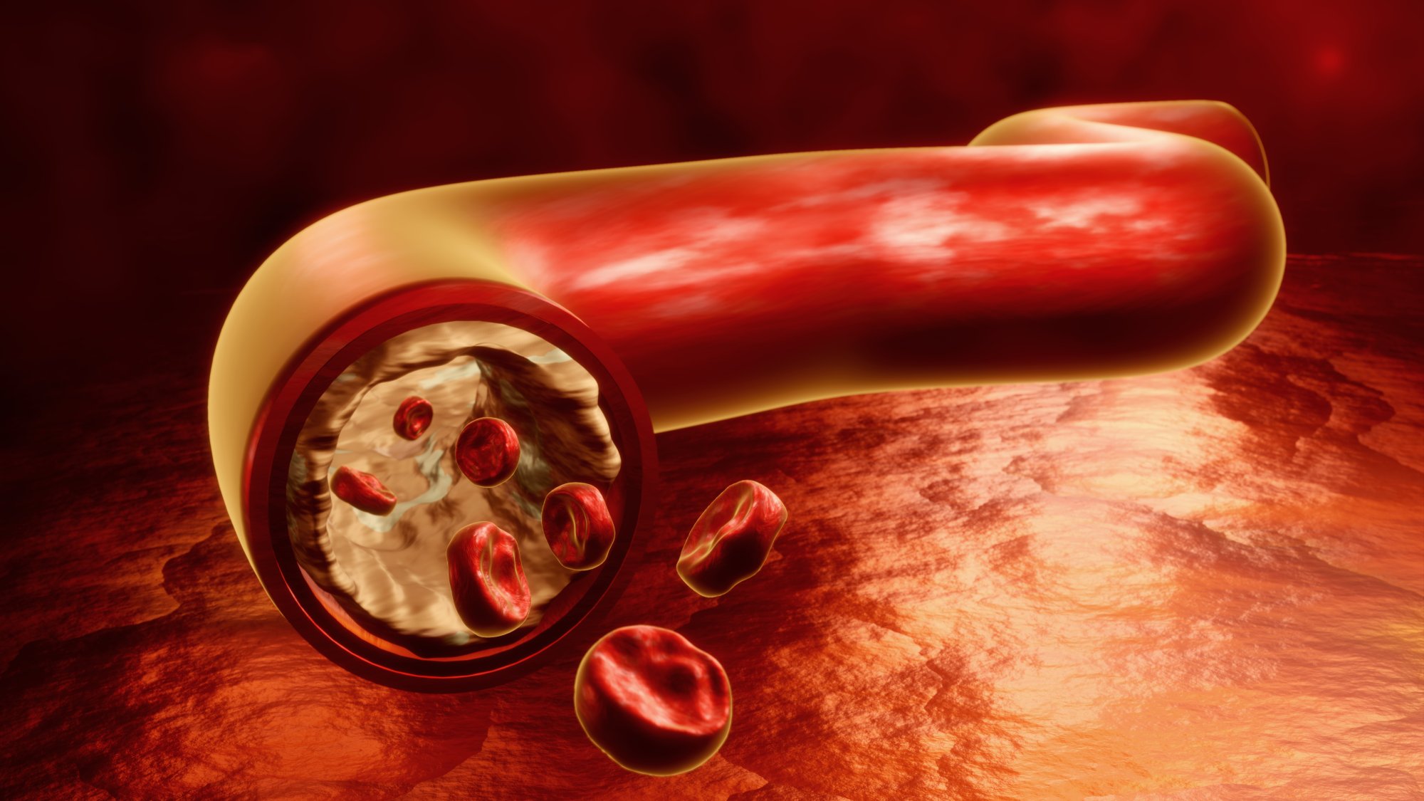
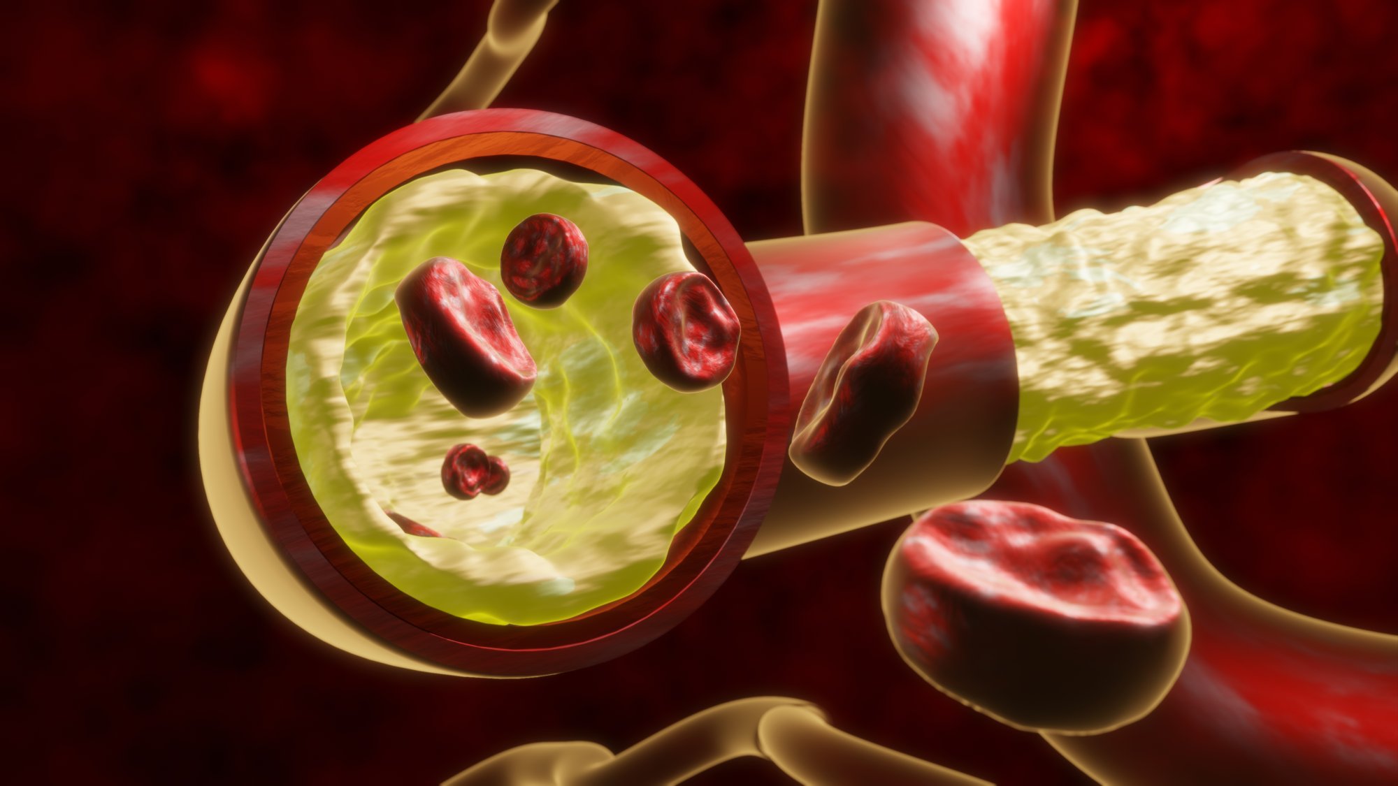
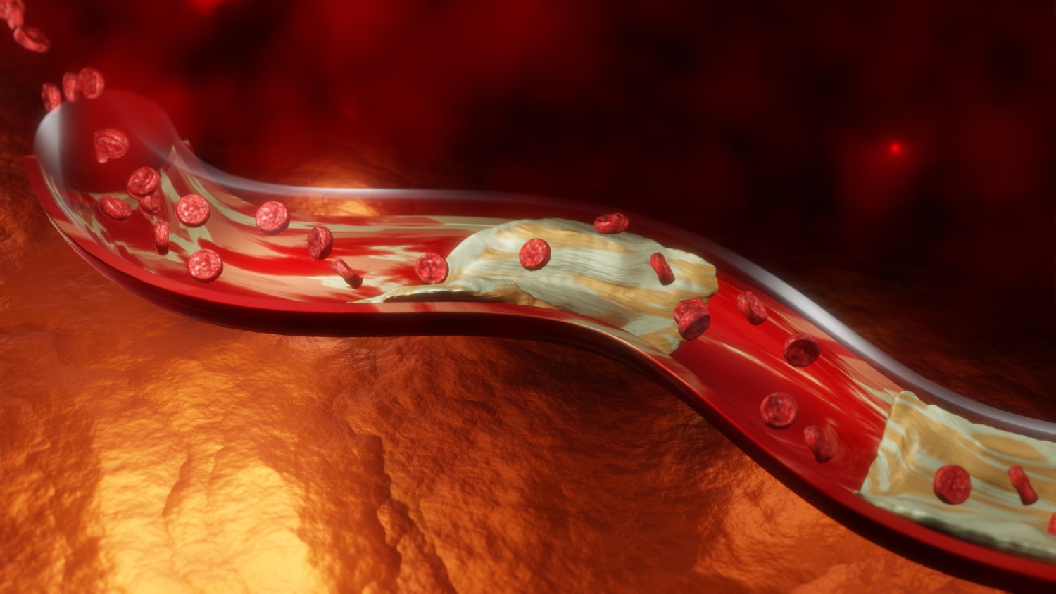
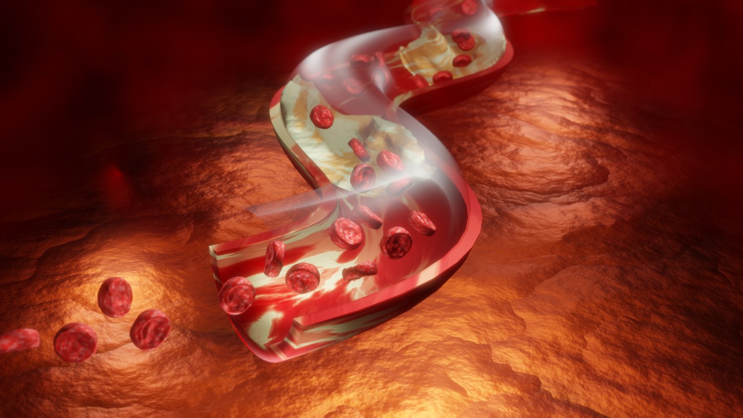
A 3D rendered illustration depicting cholesterol accumulation inside human blood vessels brings clarity to a process that usually remains invisible within the body. This visual representation transforms a microscopic event into an accessible scene by showing the interior walls of major pathways in a lifelike environment. Instead of imagining an unseen buildup, the viewer observes a realistic interpretation of how deposits develop, spread, and alter the vessel’s structure over time. This form of illustration helps learners understand the physical changes that occur in the circulatory system with remarkable accuracy.
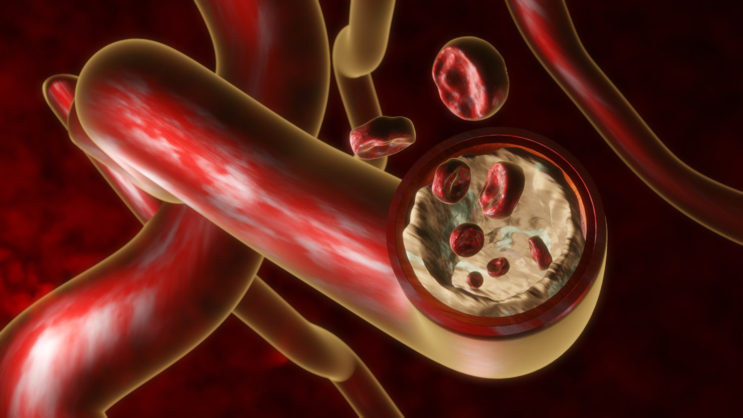
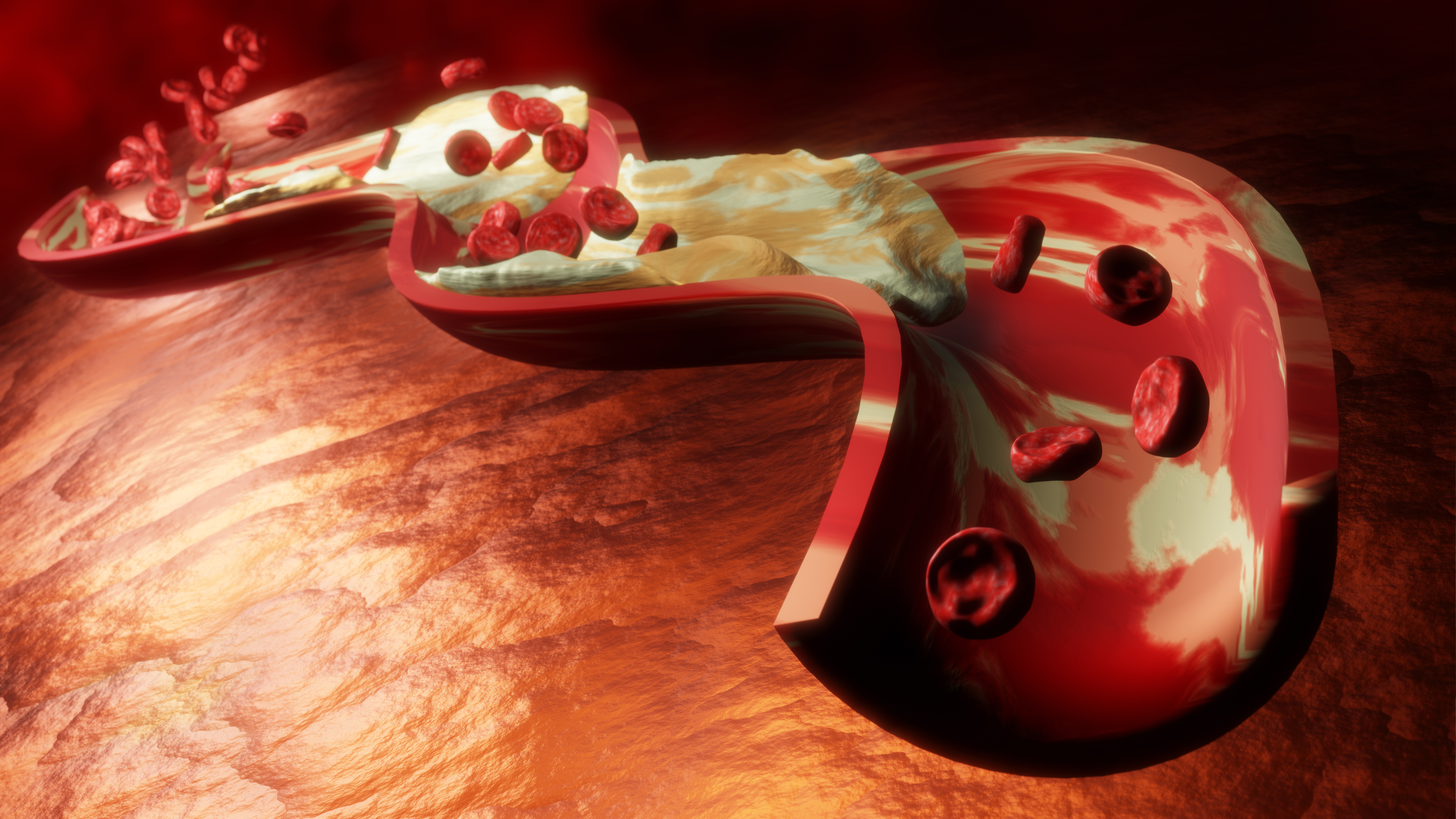
Inside a rendered vessel, the smooth internal surface appears as a uniform layer, representing the healthy condition. Gradually, raised patches of accumulated material appear along the wall. These patches vary in shape and thickness, creating a distinct contrast with the surrounding tissue. In three-dimensional space, each small mound becomes easy to recognize. Shadows and lighting enhance the textures, highlighting ridges, curves, and uneven surfaces of the buildup. The narrowing of the passage becomes visually clear as the deposits grow inward, reducing the available space for circulation.
Such images also illustrate how uneven distribution contributes to the progression of the change. Some areas may display small clusters, while others show thickened layers stretching across the vessel. The varied landscape demonstrates why movement inside the channel becomes restricted. A 3D render makes these relationships easier to understand than flat diagrams or symbolic sketches. The viewer sees how the vessel adjusts around the growing material, revealing the tension between structural stability and internal obstruction.
This rendered scene supports health education by providing a realistic reference for discussions about lifestyle choices, diet, and wellness. Many people hear about vessel blockage without truly recognizing how the process unfolds. With a detailed visual, educators can show how buildup accumulates gradually rather than forming all at once. The smooth transition from a clear pathway to a narrower one illustrates how long-term habits influence internal conditions. This helps audiences understand why prevention and early awareness matter.
In medical and anatomy training, such images play a significant role in explaining how the structure of a vessel affects circulation. Students can study the uneven surfaces, observe how deposits extend outward, and examine how the remaining opening changes shape. This knowledge is important for understanding symptoms associated with restricted flow. A 3D image allows instructors to emphasize the complexity of the landscape inside the vessel, showing that the buildup is not simply a flat patch but a multilayered arrangement with depth and texture.
Scientific communication also benefits from this type of rendering. Researchers can use detailed visuals to illustrate results, explain mechanisms, or highlight stages of progression. The ability to rotate the model, adjust lighting, or enlarge specific areas makes it easier to present information clearly in presentations, reports, or digital guides. Because the rendering remains detailed even at large scales, it works effectively in classrooms, conferences, or online platforms.
In summary, a 3D rendered depiction of cholesterol accumulation inside human blood vessels offers a clear, realistic, and highly informative view of an important biological process. By revealing the contours, textures, and gradual development of deposits, the illustration helps learners understand how vessel structure transforms over time. This combination of visual depth and scientific detail makes the rendering an effective tool for education, training, and communication across various fields.