Understanding Waxy Fat Deposits in Blood Vessels Through Detailed Visual Representation
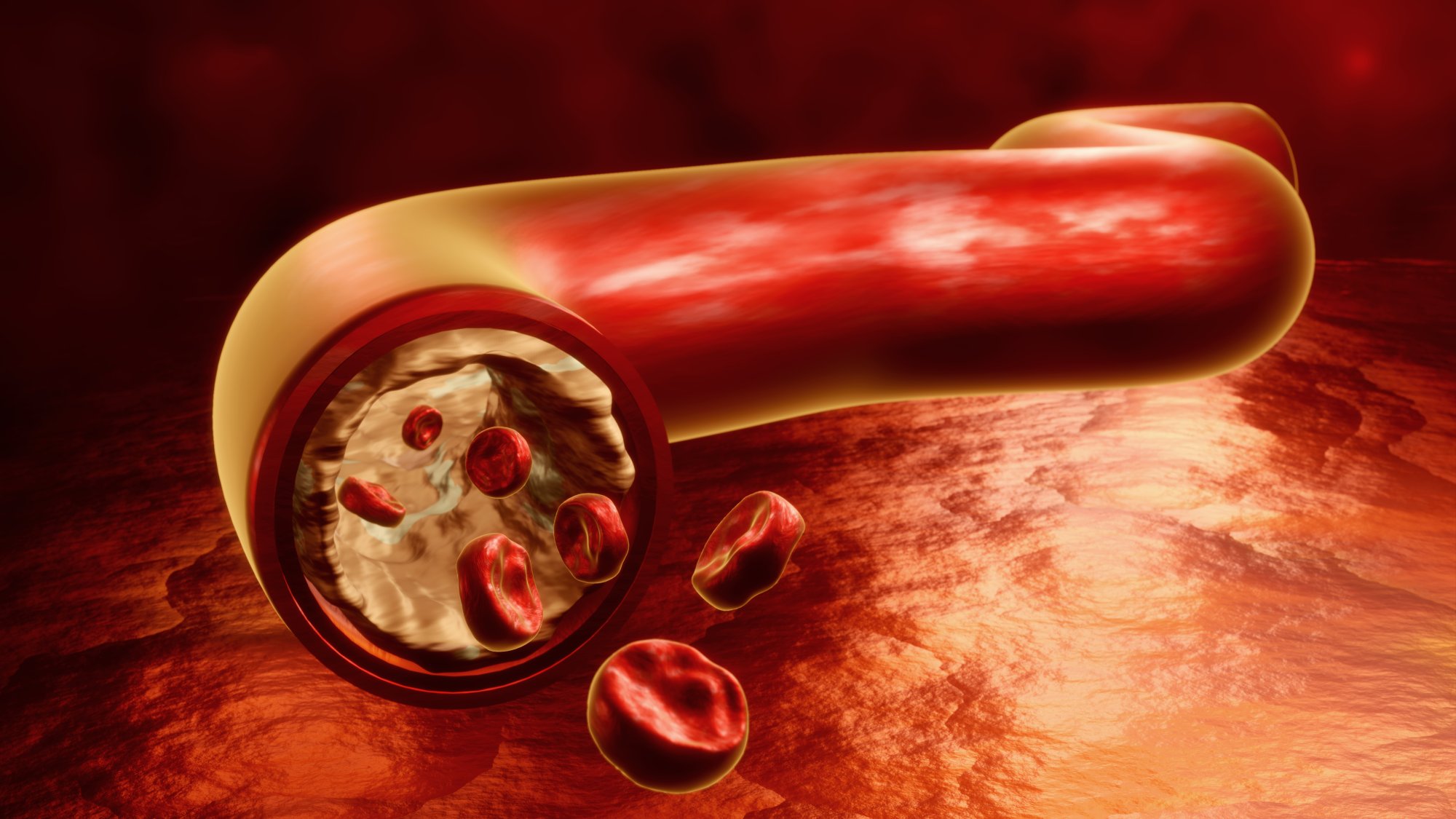
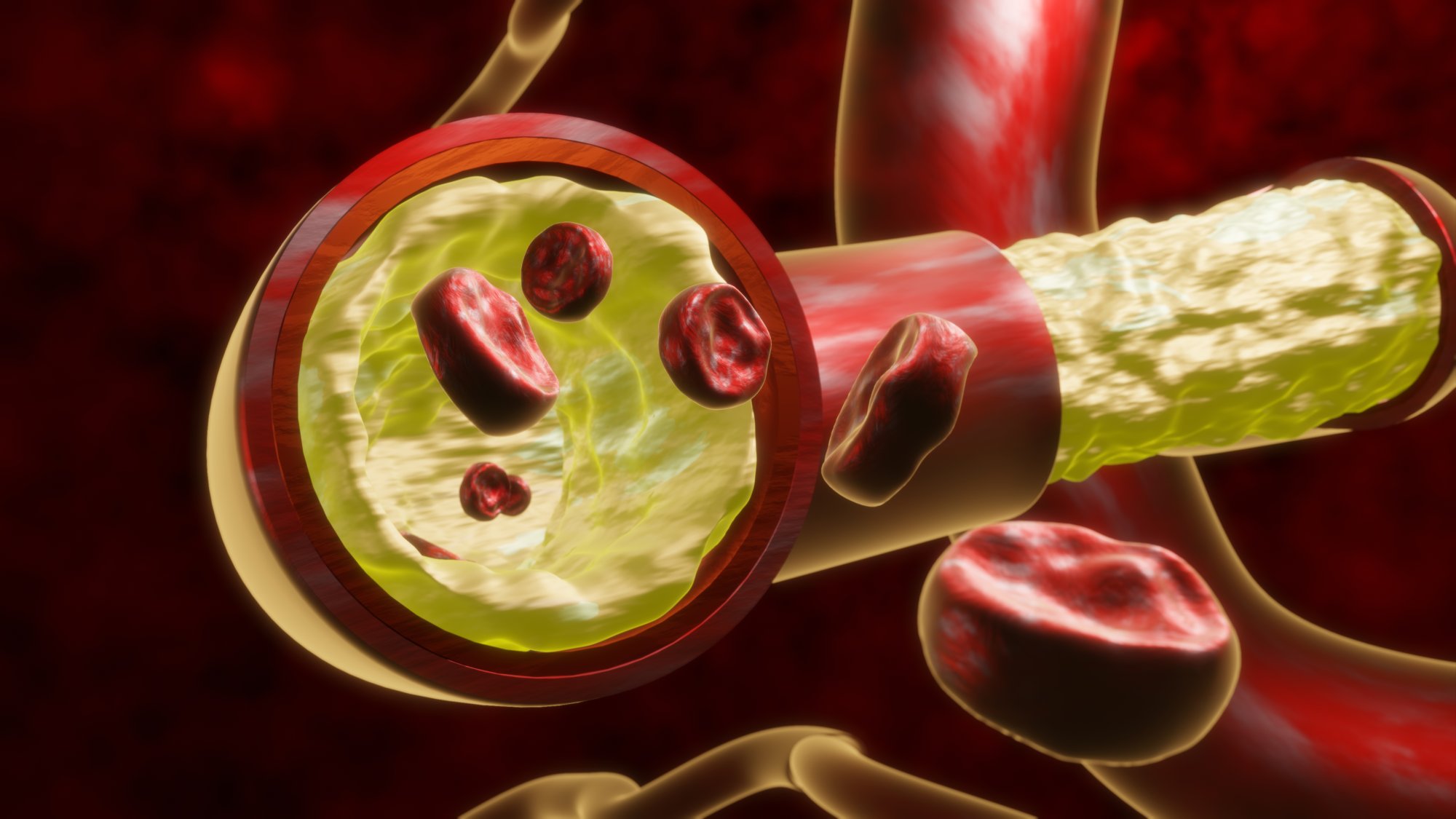
A clear understanding of waxy fat deposits inside blood vessels becomes significantly easier when presented through a precise and immersive three-dimensional illustration. These deposits, often described as soft, wax-like layers of accumulated material, gradually form within the inner lining of vessels and alter the flow of blood across critical pathways. A carefully created visual model captures the texture, volume, and distribution of this buildup, allowing learners to interpret the structural changes that occur as the material continues accumulating. Through this visual clarity, an abstract biological process transforms into something concrete and easier to comprehend.
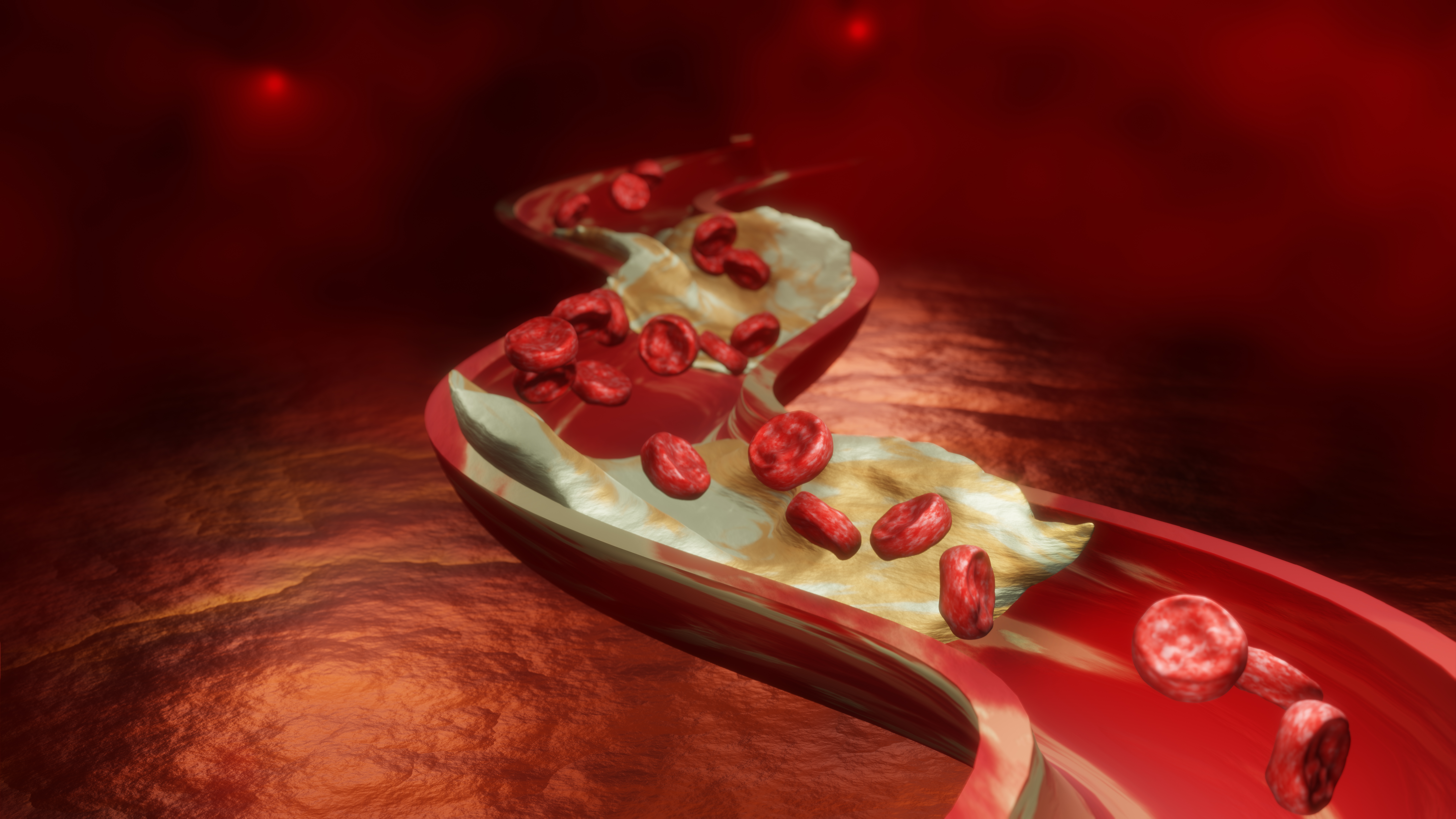
The presence of waxy fat deposits inside vessels is often associated with gradual narrowing of the channel through which blood moves. A 3D rendering showcases this narrowing by demonstrating how the internal diameter becomes constricted as layers of fatty material attach to the walls. This reduction in space alters the movement of blood, slowing the flow and increasing resistance. The rendered image highlights these shifts with balanced lighting and dimensional accuracy, making it possible to understand how a seemingly small layer of material can create notable changes in circulation. This form of illustration helps learners appreciate why early detection and awareness are crucial for maintaining vascular health.
Another important advantage of the three-dimensional visualization is its ability to demonstrate the uneven nature of the deposit. The material rarely forms a smooth surface; instead, it tends to develop in irregular clusters and layered patterns. A detailed render captures these uneven surfaces, emphasizing the textured nature of the buildup and showing how it interacts with the vessel lining. This level of detail increases understanding of how the structure evolves over time. When viewers observe the contours and positioning of the deposit, they gain insight into how the buildup disrupts the smooth passage of blood and creates turbulent flow patterns that influence overall health.
This type of visual also plays a meaningful role in understanding how the deposit progresses. A still illustration or diagram can show only a single moment in the process, while a dynamic three-dimensional representation suggests continuity and growth. The image conveys how small, scattered deposits gradually merge into thicker layers that occupy larger portions of the vessel. This progression helps explain why early stages often go unnoticed and why more advanced buildup can lead to critical consequences. The model encourages viewers to consider long-term effects, forming connections between minor initial changes and later structural challenges.
By studying a 3D illustration, viewers can also observe how the deposit interacts with surrounding components inside the vessel. Cells, fluid movement, and vessel walls all respond differently to the presence of waxy fatty material. The representation reveals how the buildup pushes against the lining, alters the curvature of the vessel, and creates zones where flow slows significantly. These details highlight both mechanical and functional effects, enriching the understanding of the entire circulatory environment. The visual structure therefore supports a more complete perspective, combining both physical and functional interpretations.
The incorporation of realistic lighting, shading, and color gradients enhances the educational impact of the rendered model. Soft textures replicate the appearance of the wax-like deposit, while contrasting tones show the difference between healthy tissue and affected areas. This artistic accuracy strengthens comprehension by helping viewers distinguish between various layers and surfaces. By drawing attention to the contrast between open channels and obstructed segments, the model encourages a careful examination of the entire system. Each visual cue adds clarity and supports deeper learning.
Additionally, such an illustration becomes a powerful communication tool for educational settings, research presentations, and medical awareness campaigns. Complex biological processes often benefit from simplified but accurate graphics, and a 3D vessel model fulfills this requirement effectively. Students, educators, and health professionals can rely on the visualization to explain conditions, illustrate progression, and highlight why prevention and timely response matter. This ability to communicate clearly across different levels of understanding makes the illustration exceptionally valuable.
In summary, a 3D depiction of waxy fat deposits inside blood vessels brings clarity to a process that is normally hidden from view. It demonstrates structural transformation, reveals spatial relationships, and shows the evolving nature of the buildup in a visually meaningful way. Through depth, detail, and conceptual accuracy, the illustration helps viewers understand how these deposits develop, how they affect circulation, and why awareness is essential for long-term vascular well-being.