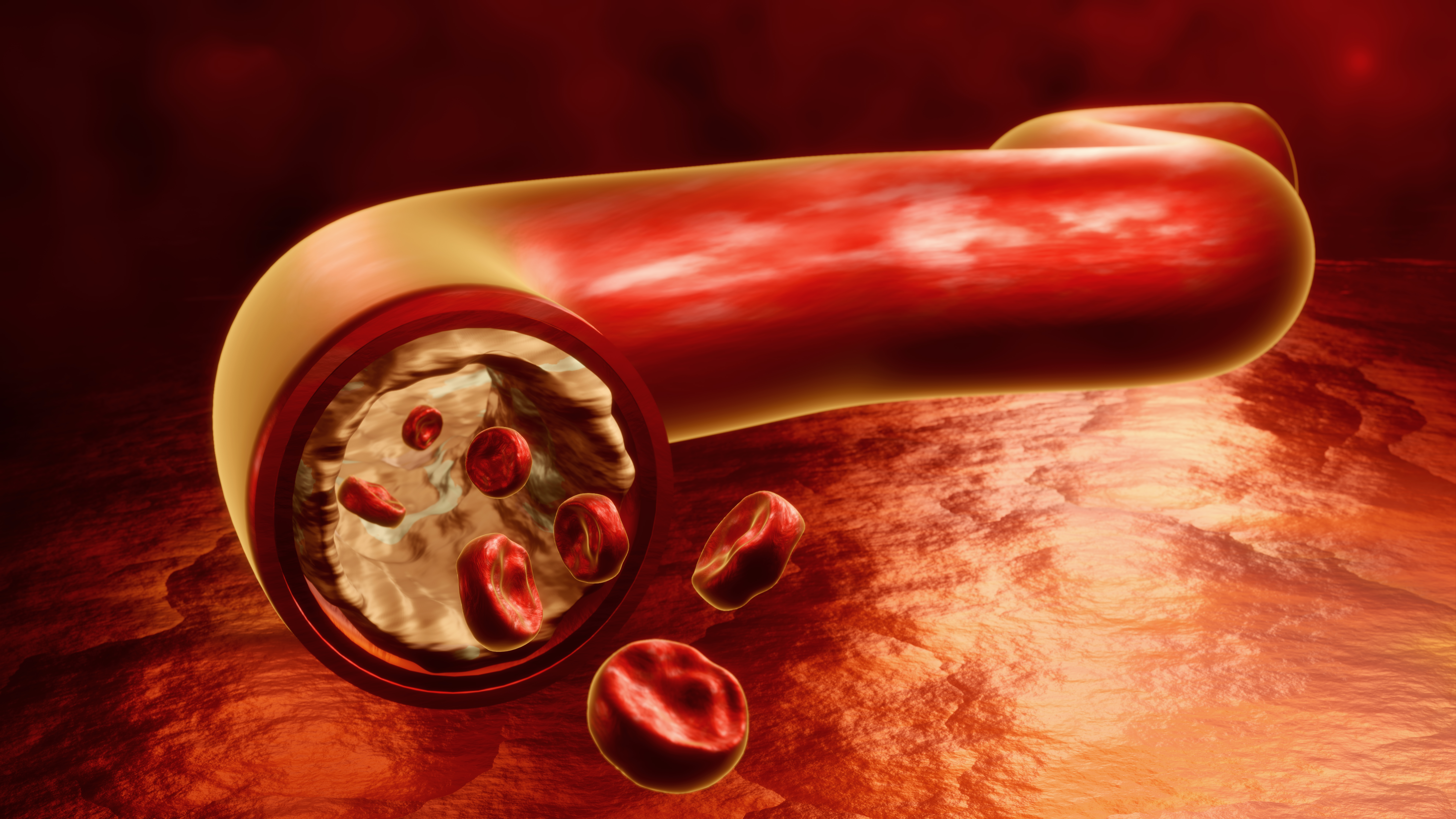
Detailed Overview of 3D Rendered Cholesterol Deposits in Human Veins and Arteries
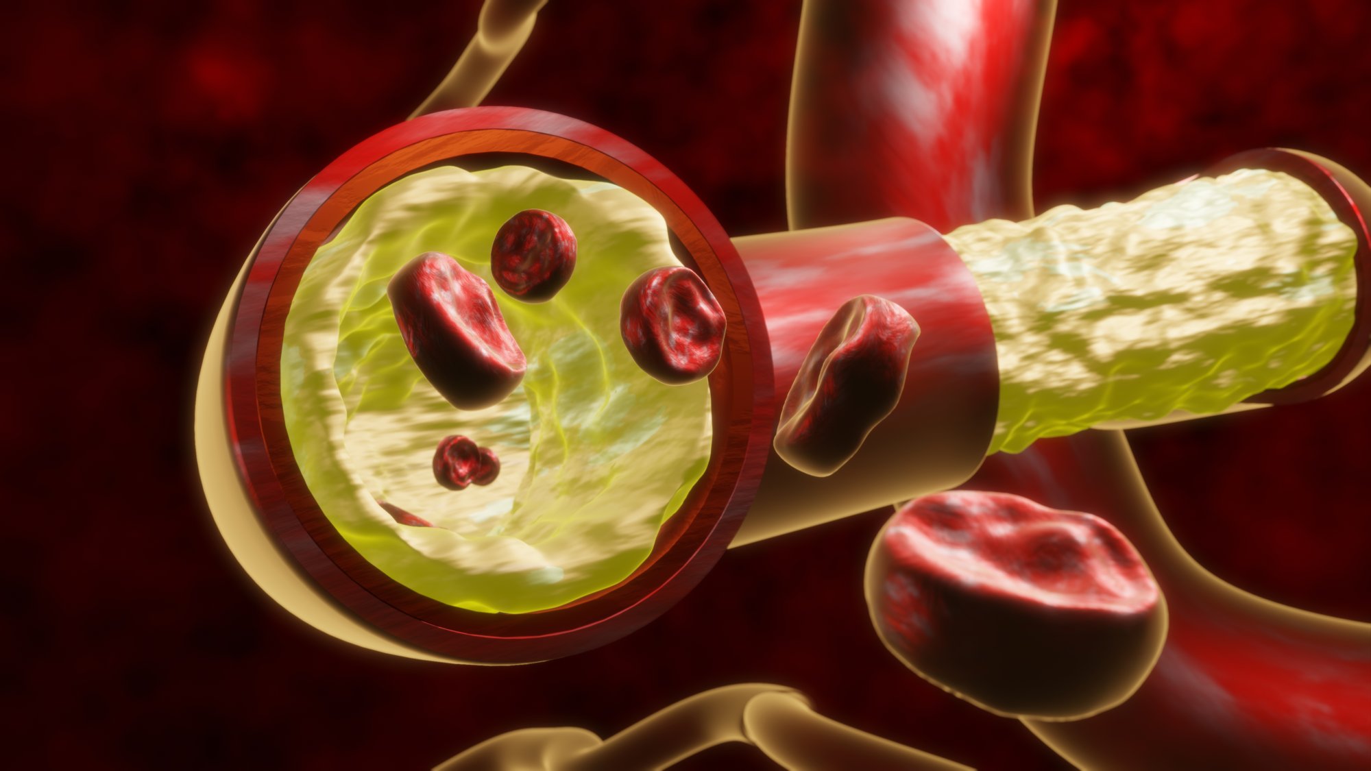
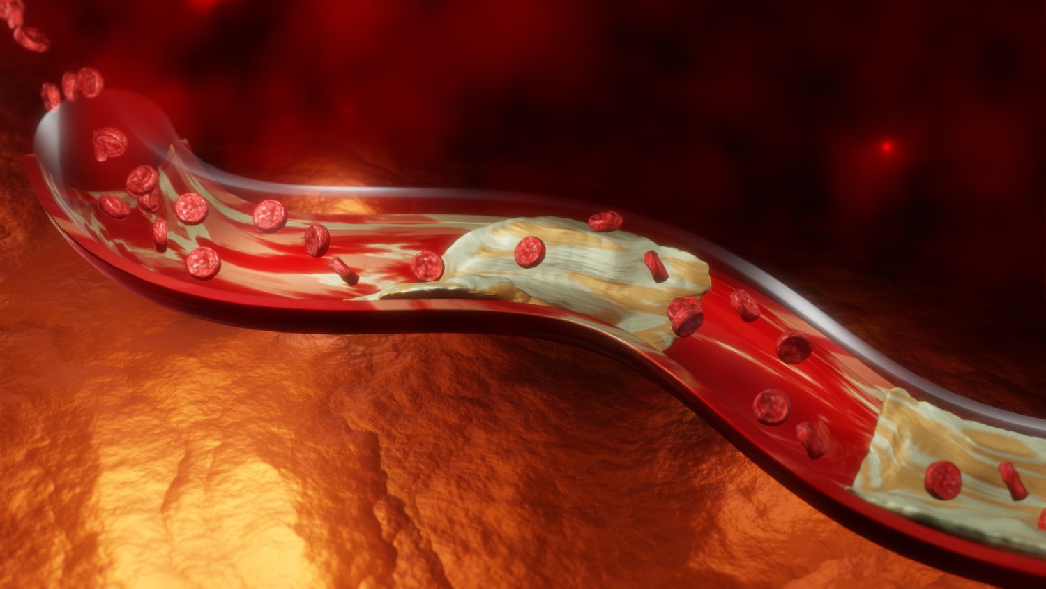
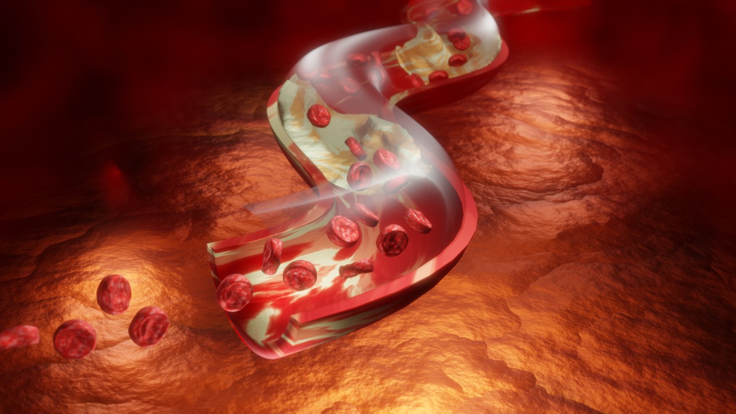
A 3D rendered illustration of cholesterol deposits within human blood vessels transforms an important biological and clinical concept into a visually clear and highly informative scene. Such an image captures the interior of major pathways that move blood through the body, revealing how small layers of material can slowly accumulate and affect flow. These illustrations allow students, health learners, and researchers to study a complex process that usually remains hidden inside the body.
Within a rendered vessel, the viewer can observe a smooth inner surface gradually altered by small clusters forming along the wall. Over time, these clusters expand, creating a raised texture that narrows the available passage. A three-dimensional image highlights this physical shift by showing depth, contours, and the irregular shapes of the developing buildup. The viewer sees how once-open channels become tighter, leaving less space for circulation. This perspective emphasizes the physical nature of the change, presenting it not as an abstract idea but as a tangible transformation.
The contrast between healthy vessel surfaces and regions with deposits is also clearer in an immersive render. Smooth red or pink walls appear uniform, while affected regions display rough, grain-like textures. Shadows and highlights strengthen the sense of space, demonstrating how buildup becomes more prominent over time. The narrowing effect becomes easy to understand even for people without a scientific background, because the reduced opening is visually apparent. The color variation helps distinguish between the normal tissue and the accumulated material.
These images support education about wellness, anatomy, and lifestyle-related risk. Many individuals hear about vessel blockage without fully understanding how the process occurs inside the body. A 3D render eliminates confusion by turning a microscopic process into a large, readable picture. Viewers grasp that the buildup does not appear suddenly; instead, it forms through gradual accumulation. This insight supports learning about prevention and reinforces the importance of choices that influence vessel health.
In professional training, such illustrations help instructors explain how the shape of a vessel changes when deposits expand. Trainees can see spatial relationships between the wall, the opening, and the raised material. These relationships affect movement of blood through the channel, making it slower or more turbulent. Understanding these changes is important for discussions regarding symptoms, potential effects, and clinical responses. The depth provided by a 3D render allows clearer observation of patterns that 2D sketches cannot fully capture.
The value of these images extends into scientific communication as well. When presented in research or medical documents, a 3D render communicates detailed information quickly. Every curve, ridge, and narrowing is represented with clarity. The structure can be rotated, enlarged, or analyzed from different angles, supporting more advanced studies. Because the image maintains accuracy even when resized, it works equally well in large posters, digital slides, or detailed guides.
In summary, a 3D rendered depiction of cholesterol deposits within arteries and veins provides a powerful visual tool for understanding an important health-related process. By displaying the inside of a vessel with depth and clarity, the illustration allows viewers to observe how deposits form, expand, and influence circulation. This clarity supports education, health awareness, clinical training, and scientific communication, making the rendered scene a valuable resource for anyone studying the human body.