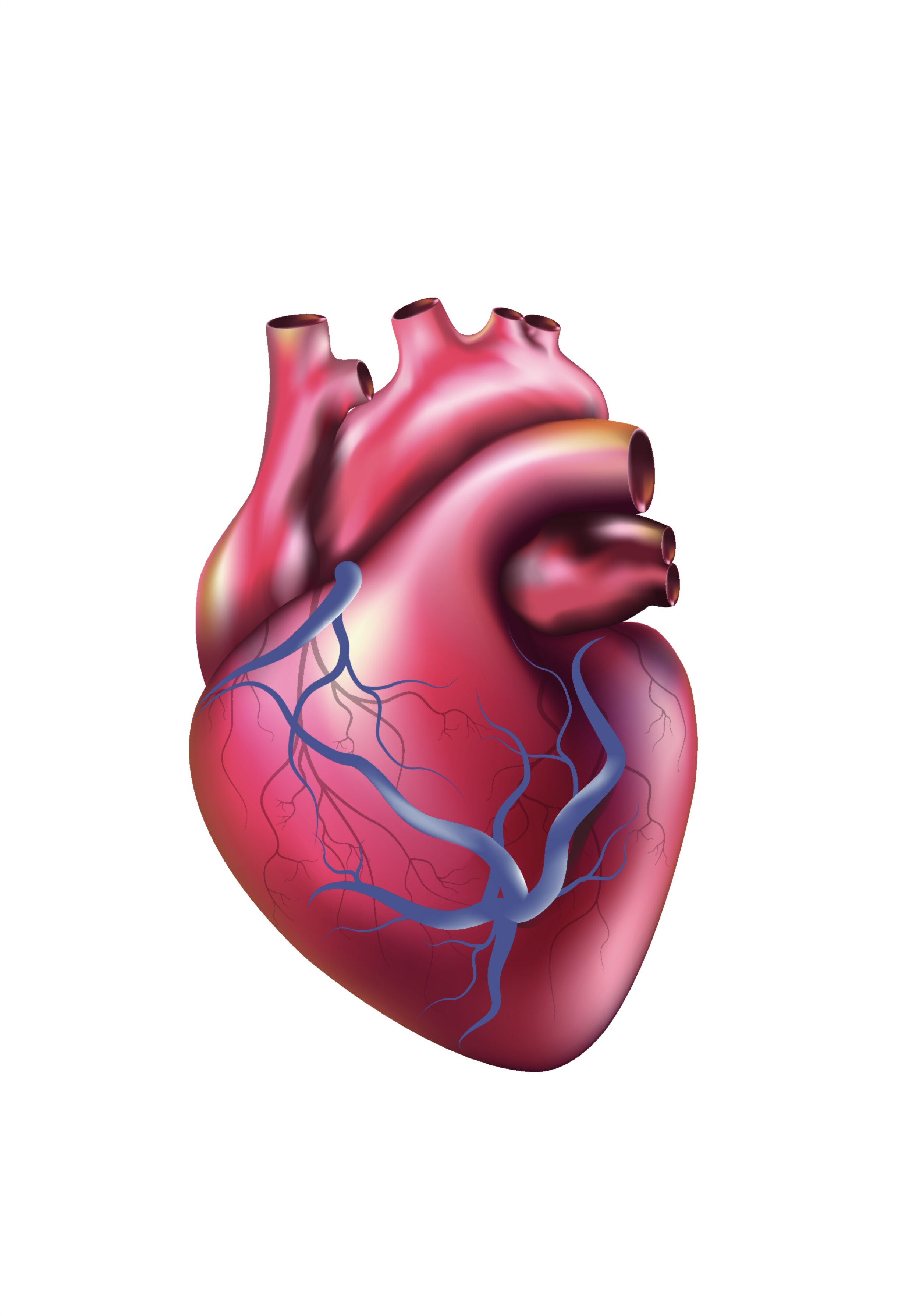
Human Heart — Structure, Function, and the Anatomical Masterpiece Behind the Rhythm of Life
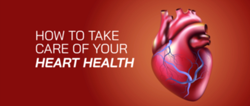
The human heart, when portrayed through a detailed vector illustration, becomes more than an anatomical graphic; it becomes a visual gateway to understanding the organ responsible for sustaining every breath, every movement, every thought, and every moment of human life. The heart is not simply a muscle hidden inside the chest but a perfectly engineered biological pump that beats tirelessly from the first weeks of development in the womb until the final moment of existence. A vector illustration of the heart captures its shape with clarity and precision, revealing its four-chambered structure, the thickness of its muscular walls, the branching of major blood vessels, and the directional flow of blood that cycles continuously through the circulatory system. Designed with smooth curved lines, layered shapes, and color-coded structural zones, the vector artwork conveys the complexity of an organ that never rests. The visual representation makes visible what cannot normally be seen: how blood enters, divides, compresses, accelerates, and exits in a coordinated sequence that defines life on a second-by-second basis.
Inside the heart, the right and left sides operate side by side, each with a unique responsibility that keeps blood moving throughout the body and lungs. In a vector illustration, this duality is often shown by using distinct colors or gradients to highlight the separation between oxygen-poor and oxygen-rich blood. The right atrium receives deoxygenated blood returning from the body and passes it into the right ventricle, which sends it through the pulmonary artery toward the lungs for gas exchange. The left atrium receives oxygenated blood from the lungs and delivers it into the left ventricle, whose powerful muscular wall propels blood into the aorta and outward to nourish every organ and tissue. The contrast in thickness between the right and left ventricles shown in the illustration reinforces that the left side must generate greater force to deliver blood throughout the body, while the right side pumps a shorter distance to the lungs. Even without words, the viewer learns that the heart is not symmetrical by accident but by necessity, with each chamber shaped by the workload it performs.
A well-rendered vector heart also highlights the network of valves that guide blood unidirectionally through the organ. The tricuspid valve lies between the right atrium and right ventricle, the pulmonary valve sits at the exit into the pulmonary artery, the mitral valve separates the left atrium from the left ventricle, and the aortic valve regulates flow from the left ventricle into the aorta. These valves are often illustrated as leaf-like structures or pointed openings that open during contraction and close during relaxation, preventing backflow and ensuring that blood advances efficiently with each heartbeat. When valves are clearly marked in the vector layout, the viewer immediately understands that the effectiveness of the heart depends not only on muscular strength but on perfect timing, coordination, and precision. The valves work as automatic gates synchronized with the cardiac cycle, creating the familiar “lub-dub” sound that represents their rapid closing and opening. Without this seamless coordination, circulation would lose its direction and the organs of the body would starve of oxygen and nutrients.
Beyond the physical movement of blood, a high-quality human heart vector illustration also makes visible the invisible system that controls the heartbeat itself — the electrical conduction network. In the graphic, this network may appear as glowing lines or highlighted pathways extending through the atria and ventricles. It typically begins at the sinoatrial (SA) node, the heart’s natural pacemaker, located in the upper right atrium. From there, impulses travel to the atrioventricular (AV) node and down the bundle fibers through the interventricular septum, eventually spreading through the Purkinje network that triggers coordinated contraction of the ventricles. The visual inclusion of these conduction routes communicates to the viewer that the heart does not require external commands to beat; instead, it generates its own electrical rhythm internally. Even when disconnected from the nervous system, the heart continues to beat through its intrinsic conduction activity. This autonomy reinforces the idea that the heart is not simply another organ but a self-governing powerhouse designed for reliability.
A vector illustration also draws attention to the major arteries and veins that form the pathways of systemic and pulmonary circulation. The vena cavae enter the right atrium, the pulmonary artery extends toward the lungs, the pulmonary veins return blood to the left atrium, and the aorta branches like a tree from the left ventricle to distribute oxygen-rich blood throughout the entire body. When depicted with smooth branching lines and emphasized gradients, these vessels become highways of life, transporting blood at variable speeds based on pressure gradients. The positioning of these vessels in the image helps the viewer trace the journey of blood and understand how a closed-loop circulatory system ensures constant delivery of essential substances. The simplicity of the visual design helps many grasp what text alone cannot explain — that every cell of the body, regardless of location, depends on the uninterrupted cycle originating within this one organ.
The vector format enhances anatomical learning by ensuring clarity at every scale. Whether enlarged on a classroom projector, printed in a medical textbook, displayed on a health awareness poster, or integrated into a digital learning module, the illustration preserves its detail. Every chamber, vessel, and pathway retains sharp boundaries and consistent proportions. The viewer can zoom into the fine lines of the coronary arteries that supply the heart muscle itself, or step back to see the organ as a whole, without any loss of resolution. This scalability reflects why vector illustrations have become central to anatomy communication: they allow the structure of the heart to be studied without distortion, making the learning experience immersive and intuitive.
The visual representation of the heart also allows connections to be drawn between anatomy and health. When arteries surrounding the heart appear narrowed or blocked in a modified illustration, the viewer can immediately infer how coronary artery disease restricts blood supply to the muscle itself. When the thickening of a ventricle appears exaggerated, the viewer recognizes the strain caused by prolonged high blood pressure. When the valves appear uneven or misshapen, the consequences of valve disorders become clearer. The illustration therefore becomes a universal language for doctors, patients, educators, and students — one that bridges scientific accuracy and emotional comprehension. Someone who has never read a medical journal can look at the image and understand the importance of keeping the heart strong because the organ’s form visually reveals its function.
Emotion also plays a subtle but undeniable role in the perceived meaning of a heart illustration. Although the anatomical heart is very different from the symbolic “heart shape” used to represent love and emotion, there is a timeless association between the heartbeat and human experience. The pulsing of the heart speeds up with excitement, slows down in moments of calm, trembles during fear, and races during joy. It responds to emotions as much as to physical needs. The vector illustration, even when purely scientific, still evokes a sense of appreciation for how deeply the heart is connected to identity, energy, affection, and the continuity of life. The viewer sees not only tissue, muscle, and chambers, but the center of everything that makes living possible.
Ultimately, a Human Heart Vector Illustration transforms invisible complexity into visible clarity. It tells the story of a perfectly organized structure that evolved to deliver nourishment to every part of the body with tireless rhythm. It highlights that the heart is both robust and vulnerable, both powerful and delicate, both mechanical and emotional. It reminds the viewer that this organ repeats its cycle without pause from before birth until the end of life, generating the pulse that defines existence. By translating anatomy into a precise and beautiful visual form, the illustration allows people to appreciate the heart not simply as a biological organ but as the central force of human survival, vitality, and connection.