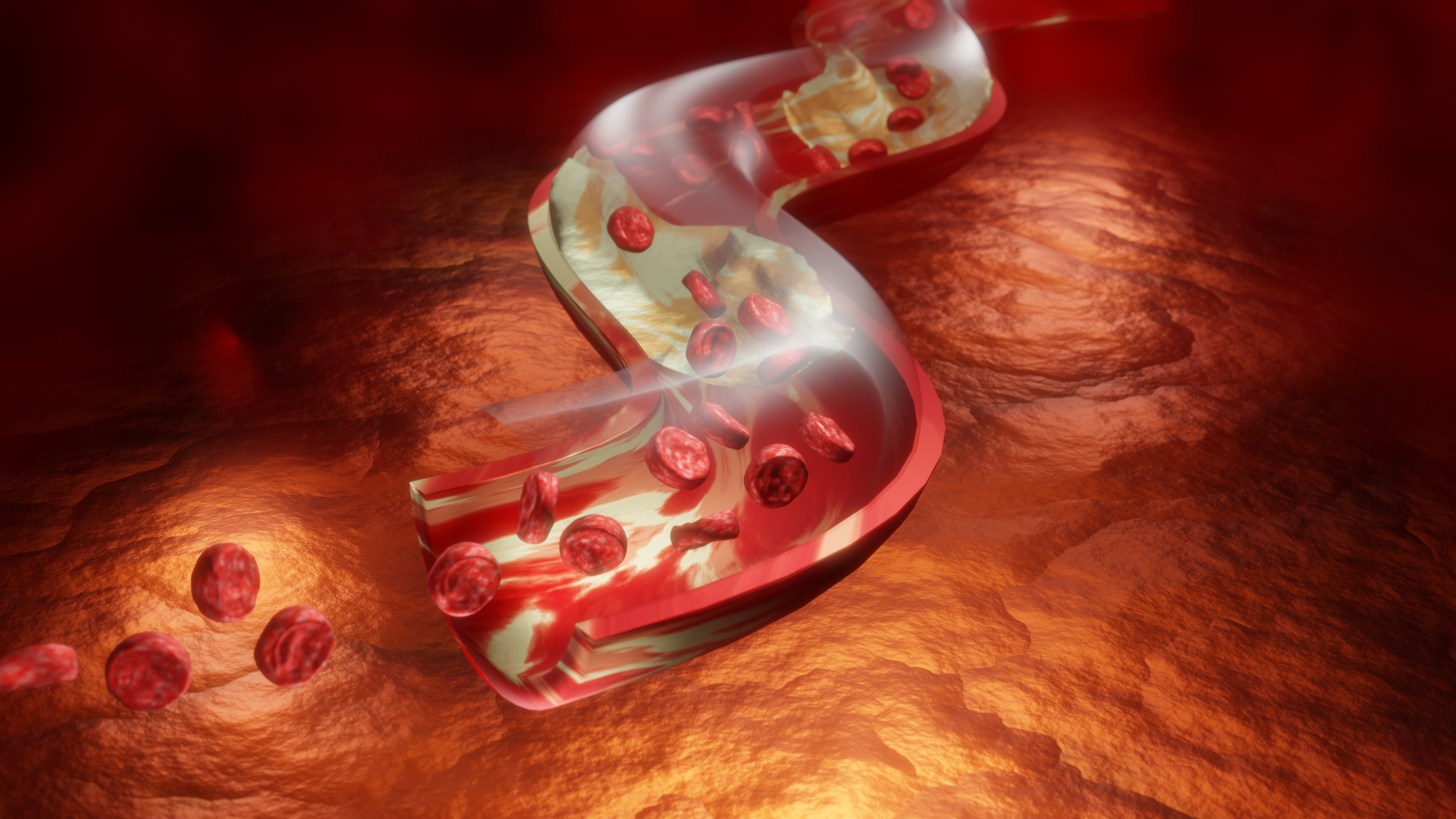
3D Rendered Cholesterol Illustration — Detailed Medical Visual Overview
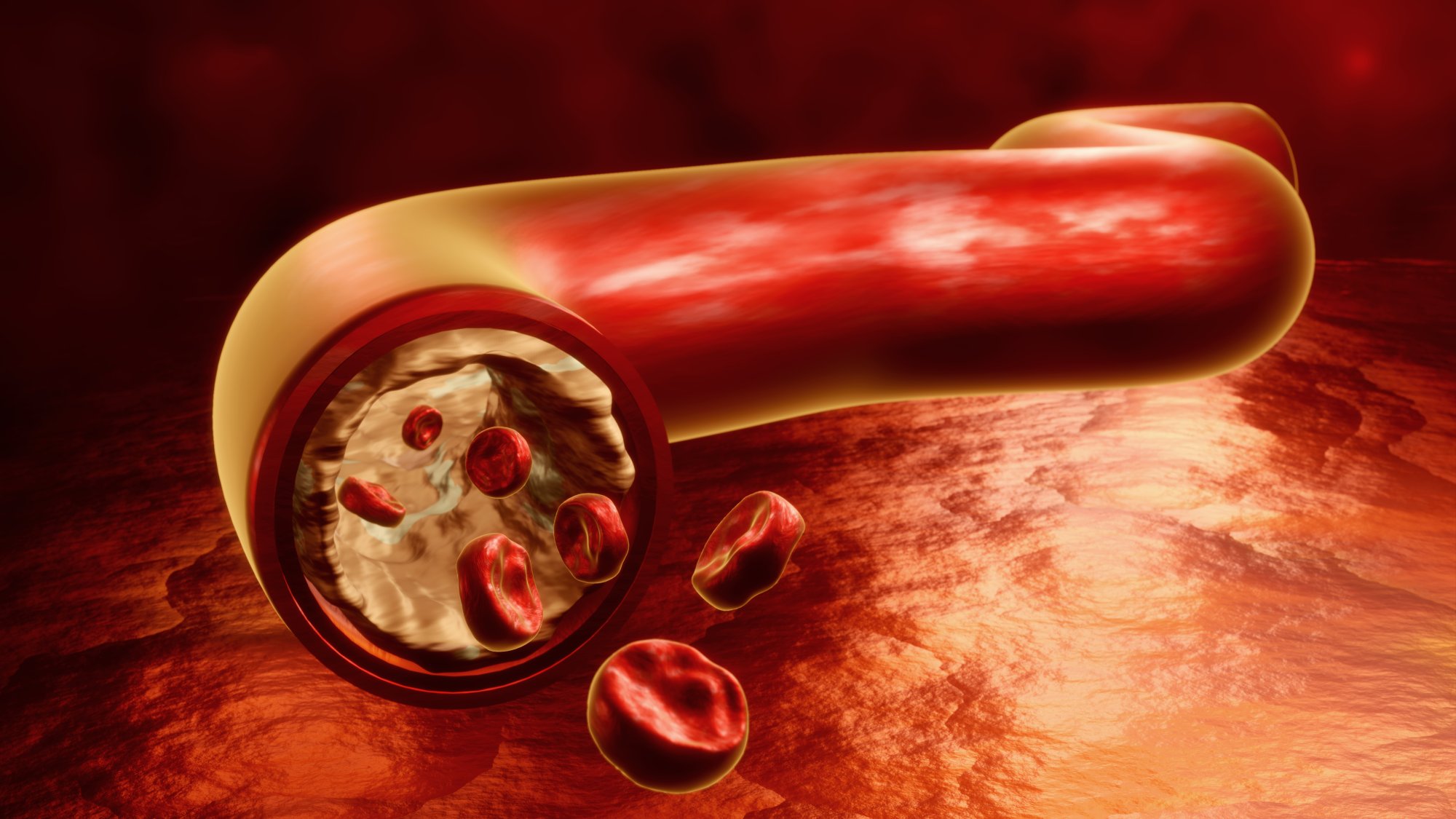
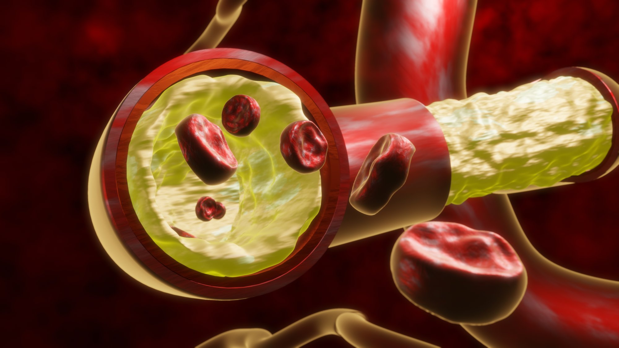
A 3D rendered cholesterol illustration provides a clear and structured view of waxy fat deposits that form inside human veins and arteries. This type of visual is especially valuable in medical learning spaces, health presentations, diagnostic discussions, and public awareness material because it converts complex biological processes into an accessible scene. The refined modeling, smooth surfaces, and layered vessel textures allow viewers to understand how these deposits accumulate and influence blood movement within the body. By presenting anatomy and biological structure in a clean, immersive form, the illustration makes an otherwise abstract process easy to grasp.
In most medical graphics, cholesterol appears as rounded clusters or soft mass formations. These groups sit along the interior walls of the vessel, showing how buildup can narrow the passageway and affect overall flow. The 3D technique highlights these deposits using detailed lighting, allowing the viewer to see how the shape and position of each cluster relates to the surrounding vessel structure. The vessel itself is often shown as a tubular form with contrasting inner and outer layers. This contrast ensures clarity when displaying where the deposits sit and how they form over time.
Another strength of this visual style is the combination of depth and surface detail. Subtle shadows and reflections help define the layers inside the artery or vein, letting the illustration explain the relationship between flowing blood and internal buildup. Such clarity is particularly important for learners who need to understand how deposits interact with the vessel lining. The smooth transitions and controlled lighting bring a realistic yet clean representation of internal structure without overwhelming viewers with unnecessary complexity. This allows the focus to stay on the buildup and its role within the system.
In educational material, this type of image helps explain discussions about body function, vessel integrity, lifestyle influence, and relevant biological reactions. Because the buildup is shown in a solid, tangible form, viewers can understand how it changes space inside the vessel. This is essential when teaching concepts related to circulation, flow restriction, or general vessel condition. Designers and instructors use these visuals in textbooks, class modules, online courses, and advisory content to show how deposits gather, how they appear within the lining, and how these formations affect movement inside the system.
The 3D format also allows adaptation for different contexts. Some variants include full vessel cutaways for broad anatomical views, while others focus on close up segments to give learners precise detail. This versatility makes the model useful in clinical explanations where professionals may need to describe how buildup changes vessel behavior. The illustration can also represent different stages of accumulation, showing viewers how the clusters appear early on and how they change shape or density as buildup progresses. These progressive visuals support guided understanding and structured learning.
In health awareness campaigns, a 3D rendered cholesterol illustration serves as a helpful tool to encourage better lifestyle choices. By showing the buildup in a realistic form, the scene communicates the seriousness of accumulation without excessive technical language. The combination of bright vessel color, smooth textures, and clearly defined cluster shapes makes the visual both memorable and educational. This helps audiences understand how routine choices influence internal buildup and overall body condition.
For digital platforms, the polished nature of a 3D illustration offers dependable compatibility with modern layouts. The clean geometry and subtle shading allow the image to appear sharp on mobile screens, tablets, or large displays. Designers can use the model in interactive guides, anatomy panels, or motion graphics where viewers explore body systems. Its clarity ensures that even when scaled or animated, the buildup retains its recognizable structure and the vessel maintains its visual integrity.
Overall, a 3D rendered cholesterol illustration provides an effective, precise, and visually appealing method to demonstrate the presence of waxy fat inside veins and arteries. Its structured design, realistic depth, and clear representation allow viewers to immediately understand the internal scene. Whether used in learning, presentation, awareness, or digital education, the illustration provides a strong foundation for understanding biological structure and the role of buildup in vessel behavior.