Detailed Muscle Structure Anatomy Showing Muscle Fibers, Myofibrils, and Sarcomere Organization
Skeletal muscle is a highly organized and dynamic tissue responsible for voluntary movement, posture maintenance, and force generation. Understanding its detailed structure, from whole muscle down to the sarcomere, is essential for physiology, anatomy, and biomedical education. A vector illustration of muscle structure typically integrates muscle fibers, myofibrils, and sarcomere organization, providing a visually intuitive representation of the hierarchical organization of muscle tissue. By combining labeled components, color-coded structures, and magnified insets, such illustrations allow learners to visualize how molecular and cellular architecture translates into functional muscle contraction.
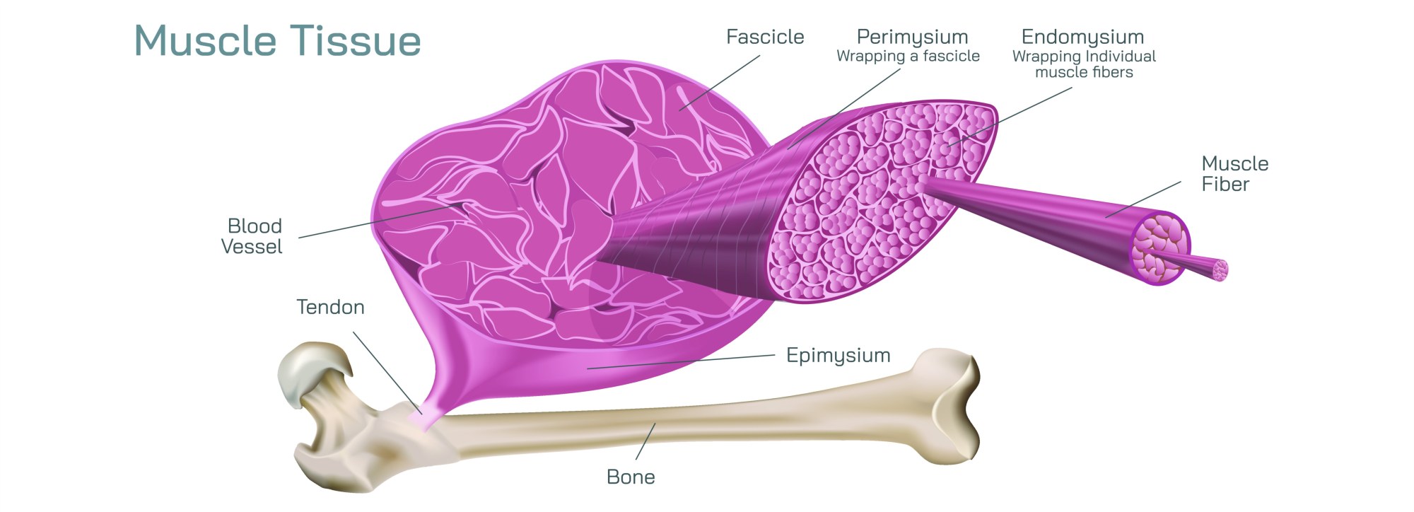
At the center of the illustration is a whole skeletal muscle, depicted with its external connective tissue layers intact. Labels identify the epimysium, the outer connective tissue sheath surrounding the entire muscle; the perimysium, which encloses bundles of muscle fibers (fascicles); and the endomysium, a fine connective tissue layer surrounding individual muscle fibers. Arrows indicate hierarchical organization, showing how fascicles aggregate to form the entire muscle and how connective tissue supports force transmission to tendons. Color coding or shading differentiates connective tissue layers from contractile tissue, providing clear visual distinction.
Muscle fibers (myocytes) are illustrated as elongated cylindrical cells within each fascicle. Labels highlight key features such as the nuclei located at the periphery, sarcolemma (cell membrane), and sarcoplasm (cytoplasm). Arrows indicate the internal arrangement of fibers and their alignment along the muscle’s longitudinal axis, emphasizing structural uniformity for coordinated contraction. Magnified insets show the cross-sectional arrangement of fibers, illustrating how densely packed fibers allow efficient force generation.
Within each muscle fiber, myofibrils are depicted as cylindrical bundles running parallel to the fiber’s long axis. Labels identify them as the contractile elements of the muscle, composed of repeating sarcomere units. Arrows indicate signal flow and mechanical force transmission along the myofibril, demonstrating how microscopic contractions sum to generate macroscopic muscle movement. Color coding differentiates myofibrils from surrounding sarcoplasm and organelles such as mitochondria, which supply ATP for contraction.
Sarcomere organization is illustrated in a magnified inset of a myofibril segment. Labels identify Z-discs, A-bands, I-bands, H-zone, and M-line, providing a clear structural map of the contractile unit. Arrows indicate the sliding filament mechanism, showing how actin (thin) filaments and myosin (thick) filaments interact during contraction. Color gradients or overlays may differentiate thick and thin filaments, highlighting the functional anatomy that enables muscle shortening and force generation. Magnified cross-sections may show overlapping filament regions, emphasizing the structural basis for the striated appearance of skeletal muscle.
The illustration may also include neuromuscular junctions, showing motor neuron axons terminating at muscle fibers. Labels indicate acetylcholine release, receptor binding, and initiation of depolarization, connecting structural anatomy with functional activation. Arrows may depict the signal pathway from the neuron to the sarcolemma and into the T-tubule system, linking excitation to contraction.
Vector diagrams often include 3D or longitudinal and cross-sectional perspectives, showing the hierarchical organization from whole muscle to fascicles, fibers, myofibrils, and sarcomeres. Color-coded layers, arrows, and magnified insets clarify relationships between levels of organization, helping learners visualize how molecular and cellular architecture supports macroscopic function. Additional features may include connective tissue integration, capillary networks, and mitochondrial localization, emphasizing support for energy supply and force transmission.
By combining muscle fibers, myofibrils, sarcomere organization, connective tissue layers, and neuromuscular components, a vector illustration of muscle structure provides a comprehensive, visually engaging understanding of skeletal muscle anatomy. Labels, directional arrows, magnified insets, and color coding allow learners to grasp the intricate architecture that underpins voluntary movement, mechanical efficiency, and force generation.
Ultimately, a detailed muscle structure vector illustration demonstrates the hierarchical integration of connective tissue, fibers, myofibrils, and sarcomeres, linking anatomical organization to physiological function. Through labeled components, hierarchical layout, magnified sarcomere insets, and neuromuscular pathways, the diagram transforms abstract muscular anatomy into an educational, visually intuitive, and engaging tool for students, researchers, and health professionals.