Human Breast Anatomy Vector Illustration Showing Structure, Lobules, Ducts, and Fat Tissue Layers
The human breast is a complex anatomical structure composed of glandular, connective, and adipose tissues, intricately organized to perform physiological functions including milk production, lactation, and hormonal response, while also contributing to secondary sexual characteristics. A vector illustration of the human breast typically emphasizes the internal architecture, highlighting lobules, ducts, adipose tissue, connective structures, and supporting vasculature, providing a comprehensive view for medical, educational, and anatomical studies. By integrating structural labeling, cross-sectional views, and tissue layers, such illustrations allow viewers to understand the spatial relationships and functional organization of the breast.
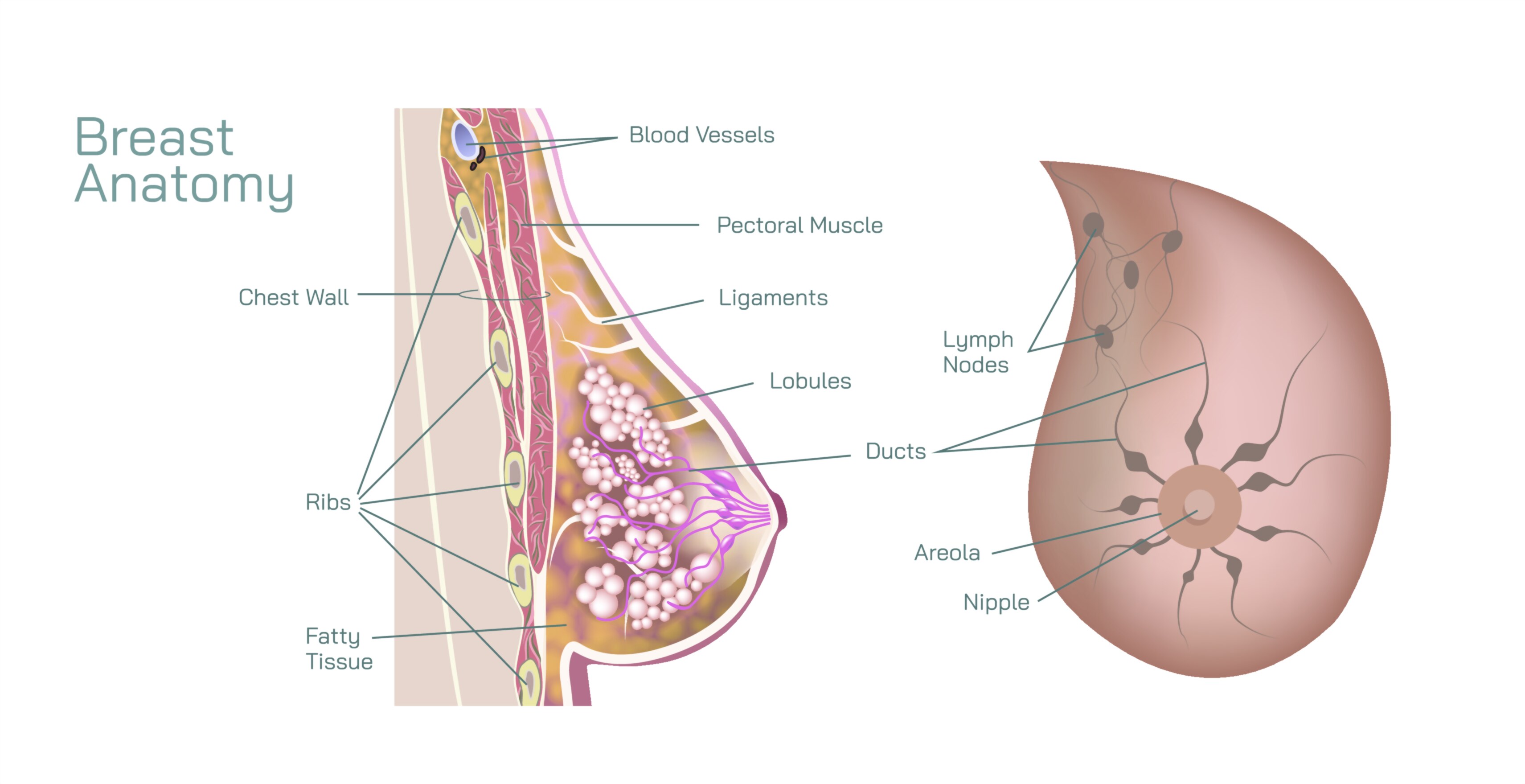
At the core of the illustration are the lobules, which are the milk-producing glands within the breast. Lobules are clusters of alveoli, which secrete milk during lactation. In vector diagrams, lobules are often depicted as small rounded units arranged throughout the breast parenchyma, color-coded to differentiate them from surrounding tissues. Arrows or labels may indicate the flow of milk from the alveoli into the ductal system, emphasizing the functional pathway that connects milk-producing structures to the nipple. Magnified insets can provide detailed views of individual lobules and alveolar arrangements, illustrating the microscopic structure that underpins milk synthesis.
The ductal system is another central component of the vector illustration. Milk from the lobules drains into interlobular ducts, which converge into lactiferous ducts, ultimately opening at the nipple. Vector diagrams often highlight these ducts as branching tubular structures, color-coded to distinguish them from lobules and connective tissue. Arrows may indicate the directional flow of milk during lactation, helping viewers visualize the continuity from glandular tissue to the exterior. The branching pattern also illustrates the functional organization, showing how numerous lobules are served by a coordinated network of ducts.
Adipose tissue is an essential component of the breast, providing bulk, shape, and cushioning. In vector illustrations, fat tissue is often depicted as shaded or colored areas surrounding the lobules and ducts, emphasizing the spatial relationship between glandular and fatty tissues. The amount of adipose tissue varies among individuals and influences the external contour of the breast. Labels may also indicate how connective tissue septa, known as Cooper’s ligaments, interweave with fat and glandular tissue to provide structural support, maintaining the breast’s shape and suspension. Shading or texturing in the diagram can differentiate fat, glandular, and connective tissues, making the layered structure immediately recognizable.
The vector illustration often includes the nipple and areola, showing their anatomical relationship to the underlying ducts and lobules. Lactiferous ducts terminate at the nipple, which serves as the exit point for milk during breastfeeding. The areola, the pigmented area surrounding the nipple, may be depicted to illustrate its structural and functional features, including Montgomery glands, which secrete lubricating fluids. Cross-sectional views in the vector diagram may reveal how ducts converge beneath the areola and terminate at openings in the nipple, providing a detailed understanding of milk delivery pathways.
Additional elements frequently depicted include blood vessels and lymphatic channels, which supply the breast with nutrients and facilitate immune surveillance. Arteries and veins may be illustrated as branching structures surrounding lobules and ducts, while lymphatic vessels are often shown draining toward axillary lymph nodes. These features are critical for understanding not only normal physiology but also clinical considerations such as breast cancer metastasis, inflammatory processes, and surgical planning. Labels and arrows clarify the course of vascular and lymphatic networks relative to glandular and adipose tissues.
Vector illustrations may also include comparative or side-by-side views of different layers of the breast. For example, a transparent overlay may show the distribution of lobules within the adipose matrix, or highlight the arrangement of ducts in relation to connective tissue. By integrating multiple perspectives—lateral, frontal, and cross-sectional—the diagram provides a three-dimensional understanding of how internal structures are organized within the breast. Arrows and shading guide the eye through the layered architecture, emphasizing both spatial arrangement and functional relationships.
Educational diagrams often annotate functional relationships, such as how hormonal signals stimulate lobules for milk production during lactation, and how the ductal system responds to facilitate milk transport. They may also depict variations in breast anatomy due to age, hormonal status, or physiological conditions such as pregnancy or lactation. Color-coded arrows can illustrate milk flow, while labels indicate tissue types, duct branches, and supporting ligaments, integrating both structural and functional aspects.
By combining lobules, ductal networks, adipose tissue, connective ligaments, and vascular structures, a vector illustration of the human breast provides a detailed and holistic view of its anatomy. It demonstrates how the various components interact to perform critical physiological functions while maintaining the structural integrity and shape of the breast. Such diagrams are invaluable in medical education, clinical training, and public health awareness, offering a clear and accessible visualization of this complex organ.
Ultimately, a vector illustration of breast anatomy conveys the interconnected architecture of glandular, adipose, and supportive tissues, emphasizing the organization from microscopic lobules to macroscopic ductal networks. Through labeling, cross-sectional views, color coding, and directional indicators, the diagram transforms abstract anatomical knowledge into a visually coherent and educational framework, enabling learners and healthcare professionals to understand both structure and function comprehensively.