Human Liver — Structure, Anatomy, Blood Supply, Functions, and Central Role in Human Metabolism
The human liver is one of the most remarkable and multifunctional organs in the human body, positioned in the upper right region of the abdomen just beneath the diaphragm, and functioning as the core metabolic hub that sustains and regulates nearly every biochemical process necessary for life. Although often depicted in simplified vector illustrations showing its outer shape and major lobes, the true complexity of the liver lies in its internal structure, the organization of its blood flow, and its vast portfolio of biochemical responsibilities. The liver is a large, wedge-shaped organ weighing approximately 1.2 to 1.6 kilograms in the average adult, making it the heaviest solid organ. Its reddish-brown appearance is a reflection of its rich vascularisation because the liver receives a dual blood supply—delivering oxygen-rich blood through the hepatic artery and nutrient-laden blood from the digestive tract through the portal vein. This dual supply uniquely positions the liver as both an endocrine responder and a chemical processor that monitors everything absorbed from food and the environment. A vector illustration of the human liver often highlights the two major lobes, the connection to the gallbladder through the biliary system, and the branching of major vessels, but beneath that surface lies an astonishing network of millions of functional units working continuously to keep the body alive.
Structurally, the liver is divided into a larger right lobe and a smaller left lobe, separated by the falciform ligament. Some anatomical models also identify two smaller lobes—the quadrate and caudate lobes—located on the underside of the liver. However, these external divisions are far less important to liver function than the microscopic internal anatomy. The true functional unit of the liver is the hepatic lobule, a hexagonal structure made up of plates of hepatocytes arranged around a central vein. Each lobule operates like an independent chemical factory, receiving mixed blood from the portal triads located at the corners of the hexagon, which contain branches of the portal vein, hepatic artery, and bile duct. Blood from these vessels flows through sinusoids—specialized capillary-like channels lined by endothelial cells and immune cells—toward the central vein. As blood passes slowly through the sinusoids, hepatocytes (the primary liver cells) extract nutrients, detoxify substances, convert molecules, synthesize proteins, and store energy molecules. Clean, processed blood drains into the central vein, moves into the hepatic veins, and eventually returns to the heart. This microscopic yet highly organized drainage system highlights the elegance of liver physiology, ensuring constant processing of metabolic, dietary, and hormonal signals.
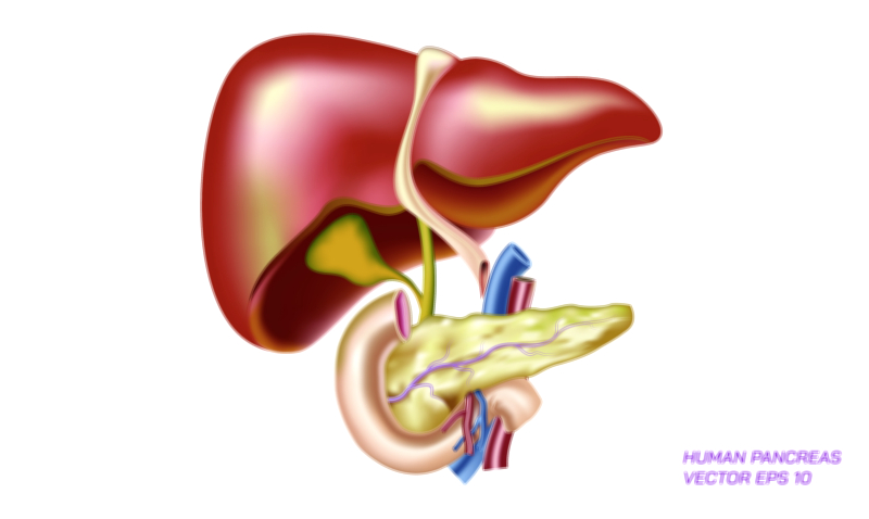
One of the distinguishing characteristics of the liver is its crucial role in digestion and nutrient handling, particularly through the production and secretion of bile. Bile is a yellow-green fluid synthesized by hepatocytes and transported through the microscopic bile canaliculi, which merge to form larger ducts that eventually drain into the common hepatic duct and then to the gallbladder for storage. When fatty foods reach the small intestine, bile is released to help emulsify fats, allowing digestive enzymes to break them down efficiently. This role not only supports digestion but also enables elimination of cholesterol, bilirubin, and toxins through the biliary tract. A vector diagram of the liver often includes the biliary tree, demonstrating how bile travels from the liver to the gallbladder and intestines, reflecting the link between digestion and liver function. Without bile formation, nutrient absorption and waste excretion would be severely impaired.
Beyond digestion, the liver performs more than 500 known functions, making it one of the broadest multitasking organs in human physiology. It regulates glucose levels through glycogen synthesis and breakdown, ensuring that the brain and muscles maintain steady energy supplies even during fasting or stress. It synthesizes crucial proteins including albumin, which maintains blood osmotic pressure, and clotting factors, which are necessary to prevent uncontrolled bleeding. It processes and breaks down hormones, drugs, alcohol, and environmental chemicals, converting them into safer compounds that can be eliminated by the kidneys or intestines. It generates cholesterol and lipoproteins required for cellular membranes and hormone synthesis, storing fat-soluble vitamins and nutrients for later release. It plays a central role in amino-acid metabolism and the removal of ammonia, a toxic byproduct of protein breakdown, converting it to urea for safe disposal. The complexity of these interconnected biochemical responsibilities explains why liver failure rapidly becomes life-threatening and why the organ is at the crossroads of so many disease processes.
Another defining attribute of the liver is its immune significance. The sinusoids contain specialized immune cells, including Kupffer cells—phagocytic cells that remove bacteria, damaged cells, and toxins from the bloodstream. This makes the liver both a metabolic factory and a powerful immune surveillance organ. It filters blood coming directly from the intestines, neutralizing pathogens before they spread to the rest of the body. At the same time, hepatocytes produce proteins of the complement system and acute-phase reactants, which contribute to immune defence and inflammation regulation. These layers of immune oversight reflect how the liver protects the body from internal and external threats while maintaining a balance between defence and tolerance so that beneficial nutrients and gut microbiota-derived molecules are not incorrectly targeted as harmful.
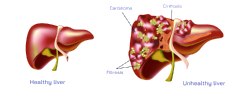
Perhaps one of the most extraordinary features of the human liver is its capacity for regeneration, unmatched by any other internal organ. When liver tissue is damaged by trauma, surgery, or disease, the remaining hepatocytes begin to proliferate in an attempt to restore lost mass. This regenerative ability allows individuals to donate up to 70% of their liver to another person and still regrow functional tissue. However, regeneration is not limitless. When chronic injury persists over many years—such as from alcohol misuse, viral hepatitis, or ongoing metabolic dysfunction—the repair process becomes distorted, producing fibrosis instead of healthy tissue and eventually leading to cirrhosis. In that unbalanced state, regeneration becomes ineffective, replacing the regenerative framework with irreversible scarring.
Vector illustrations of the liver are widely used in medical education, digital health graphics, anatomy charts, and healthcare awareness because visualizing the liver helps explain major pathways of disease, surgical procedures, and metabolic concepts. These illustrations typically show the smooth outline of the organ, major lobes, the location of the gallbladder, and key blood vessels such as the hepatic artery, portal vein, and hepatic veins. More detailed diagrams may highlight bile ducts, hepatocytes arranged in lobules, sinusoids, central veins, and portal triads—features essential to understanding how the liver filters blood and produces bile. Even simplified images give viewers a strong spatial understanding of where the liver is located and how its connections integrate digestion, metabolism, and circulation.
The centrality of the liver to human life makes the study of its structure and functions fundamental to understanding health. Whether processing nutrients, detoxifying chemicals, producing energy substrates, defending the immune system, regenerating after injury, or filtering blood, the liver is an irreplaceable biochemical powerhouse. When functioning well, it sustains a stable internal environment and allows the body to adapt to nutritional changes, environmental exposure and stress. When damaged, it disrupts multiple systems simultaneously, affecting digestion, metabolism, circulation, nervous function, immunity, and hormonal balance.
In essence, the human liver is not simply another organ—it is the metabolic command centre of the body, coordinating biochemical processes with precision and efficiency. Vector illustrations may capture its external shape and major pathways, but its inner complexity is far greater: a dense network of regenerative cells, immune sentinels, metabolic reactors, vascular channels and biochemical control systems all working in harmony. Understanding the liver’s structure and its indispensable functions reinforces why protecting liver health is one of the most essential foundations of long-term human well-being.