Human Hand Skeleton Illustration: Bones, Joints, and Structure Explained
The human hand is one of the most intricate skeletal structures in the body, and an illustration of its bones and joints reveals how strength, precision, flexibility, and coordination are achieved through an arrangement refined by evolution. When the hand is depicted schematically, its bony architecture appears almost like a branching system: a solid wrist base supporting the palm and five radiating digits. Yet behind that seemingly simple form lies a network of 27 individual bones that articulate through dozens of joints, creating the mechanical complexity required for grasping, manipulating tools, communicating gestures, and performing the fine motor tasks that distinguish human capability.
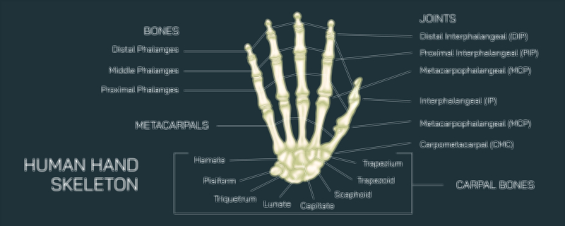
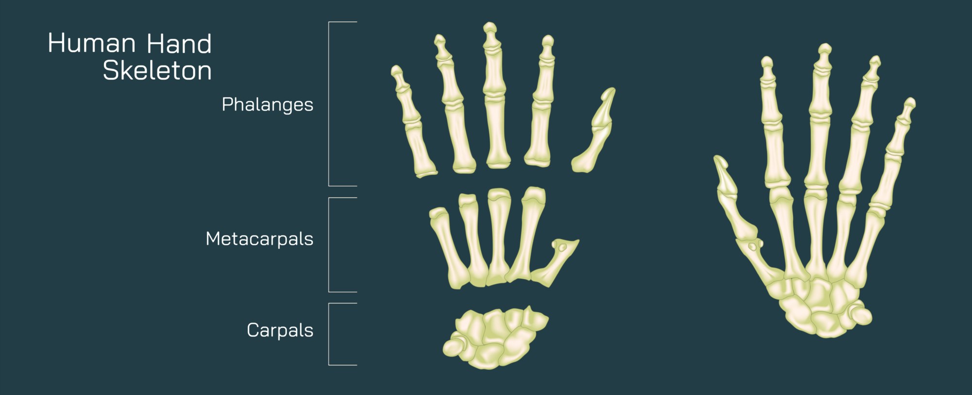
The foundation of the hand lies at the wrist, which is composed of eight small bones collectively known as the carpals. In most illustrations, these bones are arranged in two rows that form a curved arch supporting the palm. The proximal row includes the scaphoid, lunate, triquetrum, and pisiform, while the distal row includes the trapezium, trapezoid, capitate, and hamate. These carpal bones articulate with the radius of the forearm to create the wrist joint, enabling bending, extending, and side-to-side motion. Their tightly interlocked arrangement provides stability while still permitting the gliding movements necessary for wrist rotation and flexion. The carpal arch they form establishes the carpal tunnel, through which tendons and the median nerve pass—an anatomical relationship often highlighted in medical diagrams to explain conditions like carpal tunnel syndrome.
Extending from the carpal bones are the five metacarpal bones, forming the framework of the palm. Numbered from the thumb to the little finger, the metacarpals are long bones with rounded heads at the distal end that articulate with the finger bones. In illustrations, the bases of the metacarpals are shown articulating with the carpal bones to form the carpometacarpal (CMC) joints, while their distal ends connect to the finger bones through the metacarpophalangeal (MCP) joints—commonly called the knuckles. The MCP joints allow flexion, extension, abduction, and adduction, making it possible to spread the fingers and form a fist. The first metacarpal, associated with the thumb, is especially prominent in diagrams because it is oriented differently from the others; this rotational position provides the thumb with its extraordinary range of motion and contributes to the opposability that enables humans to grasp and manipulate objects with precision.
Beyond the metacarpals, the phalanges make up the skeletal structure of the fingers and thumb, and illustrations typically emphasize their number and articulation. Each of the four fingers has three phalanges: proximal, middle, and distal, while the thumb has only two: proximal and distal. This structural difference plays a major role in thumb mobility and grip function. The joints connecting these bones include the proximal interphalangeal (PIP) joints, located between the proximal and middle phalanges, and the distal interphalangeal (DIP) joints, found between the middle and distal phalanges. The thumb has only one interphalangeal (IP) joint, located between its proximal and distal phalanges. These joints collectively enable precise, independent bending of each finger segment and are the mechanical basis for gripping, pinching, typing, hand signaling, and tool use.
Hand skeleton illustrations frequently highlight the thumb carpometacarpal (CMC) joint, which is structurally unique. It is a saddle-shaped joint formed between the first metacarpal and the trapezium carpal bone, allowing a degree of rotation and opposition unlike any other finger joint. This ability to bring the thumb across the palm to touch the fingertips gives the hand its extraordinary dexterity. When shown in diagrams, this joint is often marked to explain why conditions like osteoarthritis commonly affect the thumb base due to the stresses of repeated gripping.
The alignment and arches of the hand also play a crucial role in its function. Skeletal illustrations often show two major arches: the longitudinal arch, running from the wrist up the center of the palm to the middle finger, and the transverse arch, curving across the palm near the distal row of the carpals. These arches allow the hand to conform to various shapes while maintaining structural integrity. They distribute force during gripping to prevent stress concentration on any single bone. The flexibility of the hand derives not just from movement at the fingers but from the dynamic ability of these bony arches and joints to shift slightly under load.
In expanded diagrams, the interaction between bones, joints, and supporting structures—ligaments and tendons—is often emphasized. Ligaments tightly bind bones to one another and maintain joint alignment, ensuring movement remains controlled and precise. Without ligament support, the hand’s numerous small joints would collapse under tension. Meanwhile, tendons originating in the forearm insert onto the phalanges and enable finger extension and flexion. Extensor tendons run across the back of the hand, while flexor tendons travel through the carpal tunnel and attach to the front of the fingers. Although soft tissues are not part of the skeletal system, illustrations often include faint outlines of these tendons to convey how bone and muscle work together.
Another common feature of hand-skeleton diagrams is the depiction of joint motion ranges. Arrows show that MCP joints allow bending and spreading, PIP and DIP joints primarily bend and straighten, and the thumb joints—particularly the CMC joint—permit movement in multiple planes. These arrows highlight why each class of joint has evolved to supply a specific set of motions while preserving stability. The wrist joint in these diagrams sometimes includes circular arrows showing rotation, an essential function for orienting the hand during fine tasks.
Illustrations can also show how the skeletal structure of the hand contributes to precision grip and power grip. Precision grip, such as holding a pencil, relies heavily on thumb opposition and flexion of the index and middle fingers, while power grip—like holding a hammer—uses the combined force of all metacarpals and phalanges braced against the palm arches. The bones in the little finger and ring finger shift slightly during power grip to improve force distribution. Skeleton diagrams sometimes exaggerate these subtle movements to demonstrate their importance.
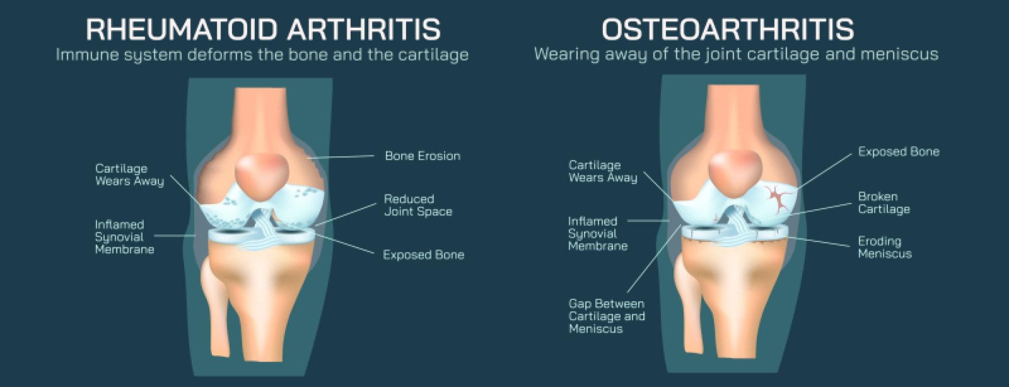
Developmental and clinical illustrations reinforce the hand’s vulnerability to injury and degeneration. Fracture diagrams highlight common injury locations such as the scaphoid in the wrist, boxer's fractures of the fifth metacarpal, and phalanx fractures from crushing injuries. Osteoarthritis diagrams show narrowing of joint space in DIP and PIP joints. Rheumatoid arthritis images highlight erosion along MCP joints and ulnar deviation of fingers when inflammation shifts joint alignment. Each of these overlays helps contextualize how the skeletal structures, though small, endure significant wear and stress.
Ultimately, a human hand skeleton illustration does more than show bones—it visualizes the architecture that enables some of the most advanced capabilities of any mammal. From the curvature of the carpals forming a protective tunnel, to the alignment of the metacarpals supporting the palm, to the multi-joint articulation of the phalanges allowing independent finger movement, every part contributes to a balance of power and precision. By translating these elements into a visual map, the illustration reveals how 27 bones work together as a coordinated system, creating the anatomical foundation for touch, craftsmanship, expression, technology, and daily function.