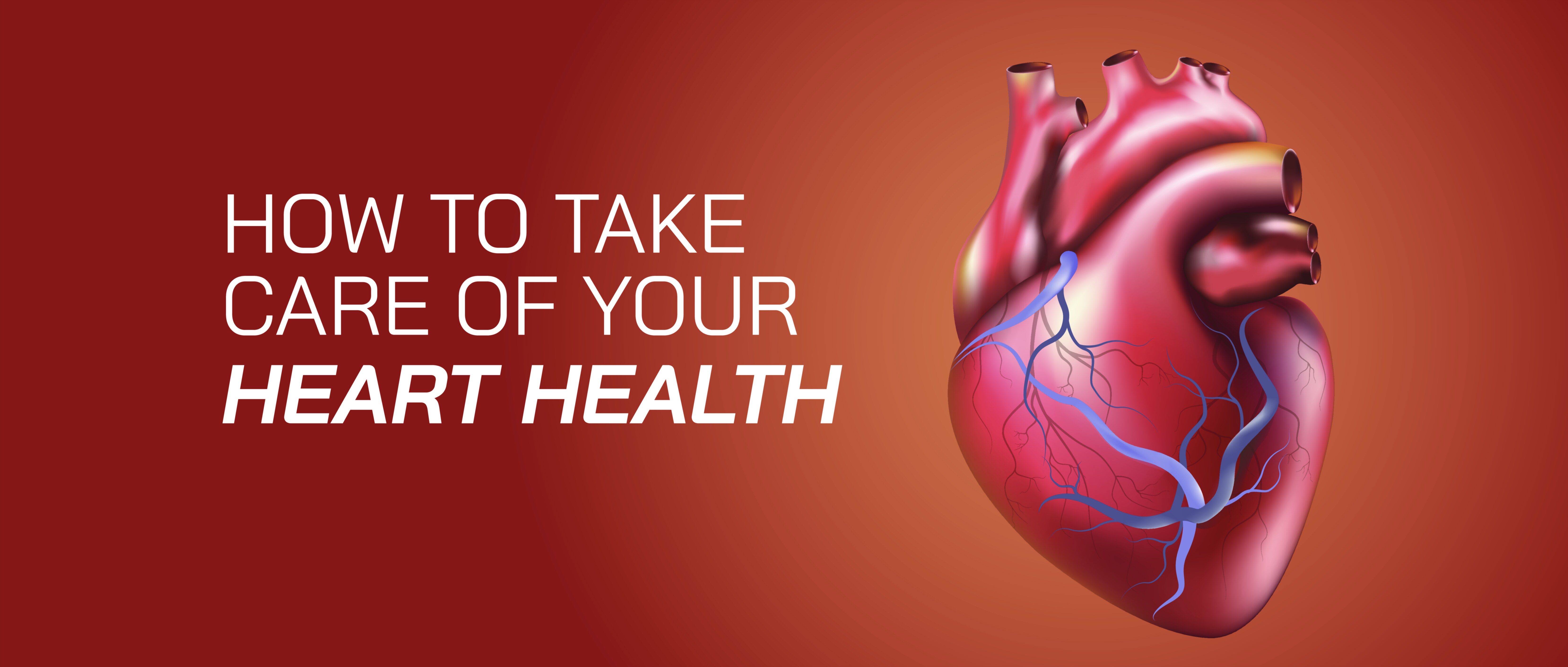
Human Heart Illustration Vector Template — Anatomical Structure, Circulatory Function, and Visual Breakdown of the Body’s Central Pump
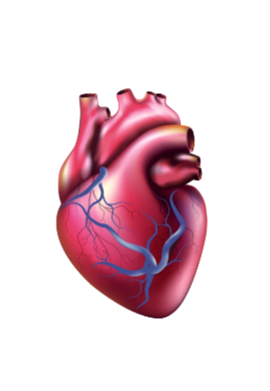
A human heart illustration rendered as a vector template represents far more than a graphic of an anatomical organ; it visually explains the biological engine that sustains life by continuously circulating blood, distributing oxygen, removing metabolic waste, and maintaining the internal balance necessary for cellular survival. The clean lines, scalable geometry, layered shapes, and sharp gradients of a vector design allow the structure of the heart to be displayed with clarity, precision, and visual hierarchy, making it suitable not only for artistic representation but for educational, clinical, and scientific materials. When viewed as a template, the illustration typically highlights the heart’s four-chambered design, depicting the right atrium and right ventricle responsible for receiving deoxygenated blood and sending it to the lungs, and the left atrium and left ventricle responsible for receiving freshly oxygenated blood and pumping it throughout the entire body. Even a simplified vector silhouette can communicate the presence of thick ventricular walls, which reflect the massive pressure needed to propel blood into systemic circulation, and the comparatively thin atrial walls designed for receiving blood entering the heart. Through shape, color segmentation, and directional flow markers, the illustration tells the story of an organ that works rhythmically and continuously, contracting and relaxing in a coordinated sequence to maintain circulation without conscious effort.
A well-designed vector heart template also communicates the directional network of major blood vessels, which play a critical role in the cardiovascular loop. The superior and inferior vena cavae enter from the body’s general circulation, delivering deoxygenated blood into the right atrium; the pulmonary artery branches from the right ventricle to send blood toward the lungs for gas exchange; the pulmonary veins return oxygen-rich blood from the lungs to the left atrium; and the large, branching aorta exits the left ventricle, distributing high-pressure oxygenated blood to tissues across the entire body. By labeling or visually differentiating these vessels through color-coded segments or contrasting pathways, the illustration demonstrates how circulation is divided into two circuits — the pulmonary circuit running between the heart and lungs, and the systemic circuit serving the rest of the body. The pulmonary circulation replenishes the blood’s oxygen supply, while the systemic circulation delivers that oxygen to muscles, organs, skin, and the brain, sustaining metabolism and energy generation. A vector template showing these dual loops creates an intuitive visual pathway, guiding the viewer’s eye seamlessly from one phase of the circulation to the next, reinforcing the idea that the heart is the hub of a looping transport system rather than a one-way conduit.
Inside the heart itself, the illustration often highlights the roles of the valves — the tricuspid, pulmonary, mitral, and aortic valves — whose alternating opening and closing ensure that blood flows forward rather than backward. The vector art emphasizes the leaf-like shapes of the valves or uses arrows to depict directional flow, helping students and observers understand that each chamber is separated by gates that open at precisely the right moment during the cardiac cycle. These valves are essential to maintaining hemodynamic efficiency: the tricuspid valve prevents backflow from the right ventricle to the right atrium, the pulmonary valve regulates flow into the pulmonary artery, the mitral valve controls flow from the left atrium into the left ventricle, and the aortic valve ensures that blood exiting the left ventricle does not return backward into the heart. When represented graphically, their positioning forms a symmetrical pattern that mirrors the heart’s rhythmic sequence of contraction, helping the viewer visualize how the organ works like an advanced hydraulic pump.
Beyond structural detail, a high-quality human heart vector template also conveys the idea of electrical control through nodes and conduction pathways that synchronize contraction. Though subtle compared to chambers and vessels, the inclusion of the sinoatrial (SA) node at the top of the right atrium, the atrioventricular (AV) node near the atrial–ventricular junction, and the branching bundle fibers running down the interventricular septum shows that the heartbeat is not random but directed by electrical impulses generated within the heart itself. These signals initiate atrial contraction first, followed by ventricular contraction, creating the coordinated pumping rhythm often associated with the classic heartbeat sound. A vector illustration can express this using directional wave curves or pulsing animation lines in digital formats, revealing how physiology depends on built-in biological circuitry that keeps the heart beating even when external systems fail.
Because the heart is central to life, a vector template of the organ has uses far beyond scientific study — it appears in healthcare branding, anatomical posters, medical infographics, surgical instruction, fitness content, first-aid education, and health awareness campaigns. The scalable nature of vector graphics ensures that the structure remains visually accurate whether displayed on a billboard or printed as a small icon on a brochure. Researchers use vector templates to annotate disease progression, showing how conditions like coronary artery narrowing, valve malfunction, myocardial infarction, or arrhythmia disrupt normal blood flow. Fitness professionals use simplified heart graphics to explain resting pulse, target training heart rate, and cardiovascular endurance. Health educators rely on the image to teach students how circulation delivers oxygen and nutrients to every cell. Clinical professionals use detailed versions to prepare patients for procedures such as angioplasty, bypass surgery, ablation therapy, or valve replacement. The adaptability of the vector template makes it an essential tool in diverse fields where clarity and anatomical accuracy are necessary.
A vector illustration of the heart also emphasizes the extraordinary workload the organ endures. Without rest, the heart beats continuously — roughly 100,000 times per day — maintaining pressure and volume cycles so that blood flows through approximately 100,000 km of vessels in the human body. The left ventricle’s muscular wall appears thicker in the graphic because it must generate sufficient force to push blood through this vast network, while the right side appears proportionally slimmer because it pumps blood only to the nearby lungs. The bilateral symmetry displayed in the vector art communicates a fundamental truth: the heart evolved not just for function but for efficiency, allocating structural thickness and orientation according to the demands of circulation. Observers can trace the flow within the template visually — deoxygenated blood entering on the right, moving to the lungs, returning oxygenated on the left, and being propelled outward — forming a cycle that repeats without interruption throughout every moment of life.
A template centered on the heart also captures the emotional and cultural significance of this organ, even though the anatomical heart differs from the symbolic “heart” shape used in art and media. Across civilizations, the heart has been associated with emotion, love, courage, and identity because people instinctively connect the rhythmic sensation of the heartbeat to feelings of intensity, excitement, and vulnerability. A technically precise vector illustration bridges the gap between scientific reality and cultural symbolism, reminding the viewer that underneath metaphor and poetry lies a true biological marvel — a muscular organ powering consciousness and experience by continuously supporting the brain and the body.
In its fullest educational role, a Human Heart Illustration Vector Template transforms anatomy into understanding. It does not merely depict chambers and vessels visually; it communicates how the heart sustains life by coordinating structure, pressure, valves, and electrical signals into a seamless cycle of pumping motion. It reminds the viewer that every breath, every movement, every thought depends on a tireless organ performing with perfect timing. Through its scalable, precision-focused design, the template makes it possible for researchers, educators, medical professionals, and learners to explore the heart not as a mystery but as a comprehensible and beautifully engineered structure. By turning biology into visual clarity, the illustration strengthens scientific literacy and deepens appreciation for the organ responsible for the rhythm of human life.