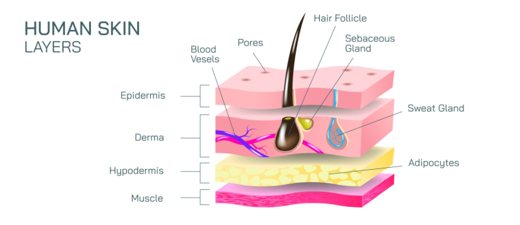
Cross Section of Red Blood Cell Structure and Function Diagram
A cross-section of a red blood cell reveals far more than a simple disk-shaped container for oxygen; it exposes a highly specialized biological design that has been refined through millions of years of evolution for efficient circulation, gas transport, and metabolic endurance. When examined closely, a red blood cell does not resemble a typical spherical cell with a nucleus and organelles. Instead, it displays a smooth, flexible, biconcave structure that resembles a doughnut with the center gently depressed rather than punctured. This shape, clearly visible when the cell is cut through its middle, dramatically increases the surface-area-to-volume ratio, allowing gases to diffuse rapidly across the membrane while maintaining structural strength. In cross-section, the central indentation appears thinner than the broader outer rim, and this variation in thickness is not accidental. The thin center minimizes diffusion distance for oxygen and carbon dioxide, while the thicker edges provide mechanical reinforcement so the cell can sustain turbulent flow through arteries and squeeze through microscopic capillaries that are narrower than the cell itself. When visualizing such a section diagrammatically, the scale of these features becomes evident, emphasizing that every contour of the cell contributes to its primary function: transporting oxygen from the lungs to body tissues and returning carbon dioxide for removal.
Inside the red blood cell, a cross-section would not show the components normally found in other cell types—no nucleus, no mitochondria, and no endoplasmic reticulum. Instead, it reveals densely packed hemoglobin molecules filling the cell like an internal suspension that dominates its volume. Hemoglobin, the protein responsible for the cell’s characteristic red color, binds oxygen in high-oxygen environments such as the lungs and releases it in low-oxygen conditions within tissues. In cross-section, this hemoglobin network appears as a homogeneous internal domain rather than a complex compartmental arrangement, demonstrating that the entire internal space of the cell is optimized for gas carriage. The absence of organelles is a defining feature, for the elimination of the nucleus during development frees up additional space for hemoglobin and reduces the cell’s metabolic demand, allowing it to rely on anaerobic energy pathways rather than consuming the oxygen it is supposed to deliver to tissues. The cross-section thus shows a cell emptied of all unnecessary components and purified into a streamlined biological vehicle centered entirely on transport.
Surrounding this interior lies the cell membrane, which forms the outer boundary visible in any structural representation. This membrane is composed of a lipid bilayer supported by a subsurface protein-based cytoskeleton that gives the red blood cell its remarkable flexibility. In a cut view, the membrane appears as a thin but continuous band around the cell’s perimeter. Although seemingly delicate, this layer is highly resilient and capable of withstanding immense physical stress as red blood cells repeatedly bend, elongate, and twist while navigating the circulatory network. The membrane also houses integral proteins that maintain ionic balance, regulate the cell’s shape, and support surface markers crucial for blood type identification. These surface antigens, although not individually illustrated in a simple cross-section, hold profound medical significance because they determine how a person’s immune system identifies red cells and whether transfusions are compatible between individuals. The continuity and uniform thickness of the membrane, as seen in a diagram, symbolize the barrier that ensures the cell’s chemical stability even as it undergoes repeated deformation in circulation.
A realistic cross-section not only displays the structure but also hints at the dynamic behavior of red blood cells within the cardiovascular system. As the body pumps blood with every heartbeat, billions of red blood cells circulate effortlessly, folding and unfolding like microscopic fabric. Their structure is so well adapted to movement through capillaries that even when compressed to half their resting diameter, they spring back to their original form without damage. This resilience comes from the cytoskeleton beneath the membrane, which appears in a cross-section as a subtle but reinforcing scaffold, maintaining the cell’s architecture. Such elasticity is essential because if red blood cells were rigid like many other cells, they would block the microcirculation pathways that sustain organs with nutrients and oxygen. Cross-sectional diagrams help highlight this balance of thinness, flexibility, and strength that makes continuous circulation possible.
In terms of physiological function, a cross-sectional illustration supports the understanding that oxygen exchange occurs across the membrane and into the hemoglobin-rich interior rather than through specialized organelles. In the lungs, oxygen diffuses across the membrane and immediately binds to iron centers within hemoglobin molecules. As blood flows to tissues, the oxygen unbinds when hemoglobin encounters low-oxygen environments and higher levels of carbon dioxide. The red blood cell then helps transport carbon dioxide back to the lungs, not only dissolved in plasma but also through biochemical conversion into bicarbonate inside the cell and partial binding to hemoglobin itself. A cross-section communicates this exchange visually by emphasizing the proximity of hemoglobin to the membrane and showing that diffusion occurs directly into the internal space of the cell rather than through compartmental channels. The cell becomes both container and active participant in gas exchange.
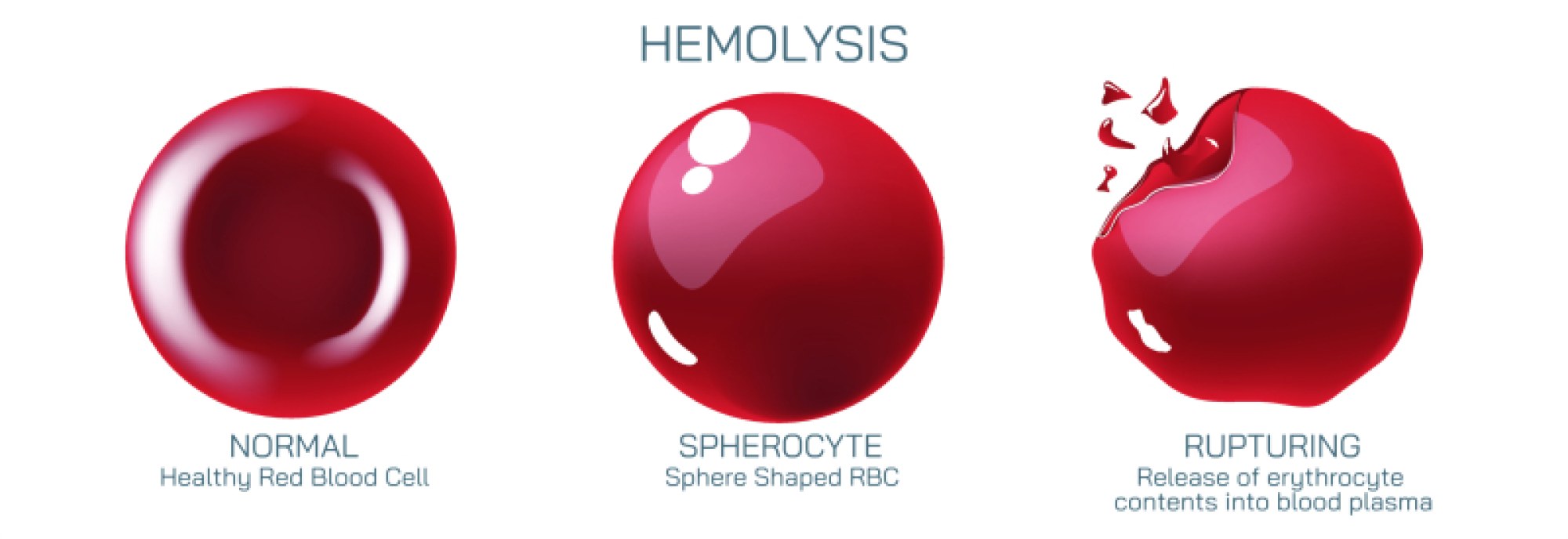
As red blood cells age—typically over 120 days—their membranes gradually become less flexible, their cytoskeleton stiffens, and their ability to navigate the narrowest capillaries declines. The spleen acts as a filtration organ that identifies and removes aging or damaged red cells from the bloodstream. A cross-section of a healthy red blood cell contrasts sharply with that of a deteriorated one: the smooth curvature and strong membrane seen in youthful cells eventually give way to irregularities and fragility. These structural changes underline why constant production of new red cells in the bone marrow is essential and why diseases that alter the shape of the cell, such as sickle cell anemia, lead to severe medical consequences. When the cell shape deviates from its biconcave design, the function breaks down, capillaries become obstructed, tissues become starved of oxygen, and circulation loses its reliability. In this sense, structure not only defines function—it preserves life.
A cross-section of a red blood cell therefore becomes a window into the harmony of biological form and purpose. The outer membrane seals the cargo and enables elasticity, the biconcave shape maximizes surface area and optimizes diffusion, and the filling of hemoglobin molecules transforms the cell into a system of oxygen delivery. Everything unnecessary has been removed so that what remains can serve a singular function with remarkable endurance. Observing the cell in cross-section does more than illustrate anatomy; it reveals the logic of efficiency that governs living systems. The red blood cell is not just a carrier of oxygen but an embodiment of biological engineering, showing how structure, composition, and behavior unite to support the most fundamental requirement of human survival: the continuous delivery of oxygen to tissues and the return of carbon dioxide for removal. Through this lens, the cross-section becomes not only a diagram but a vivid reminder of the precision underlying life at its smallest and most essential scale.