Types of Muscles: Skeletal, Smooth, and Cardiac Muscle Structure Explained
Muscles are the biological engines of movement, posture, internal regulation, and life-sustaining motion, and although they all perform work through contraction, their structure and specialization differ so profoundly that the human body relies on three separate muscular forms: skeletal, smooth, and cardiac muscle. Understanding these muscle types in a continuous explanation reveals how the body synchronizes voluntary control, involuntary motion, and rhythmic autonomic activity across every organ system. Even though each muscle type relies on actin and myosin proteins sliding across one another to generate force, the way they are organized, triggered, and coordinated varies dramatically depending on the organ they serve. When examined comparatively, skeletal muscle appears as a precisely aligned tissue designed for powerful and voluntary body movements; smooth muscle emerges as the adaptable operator of internal organs functioning without conscious involvement; and cardiac muscle stands uniquely between them, blending structural strength with automatic rhythmic contraction that sustains life.
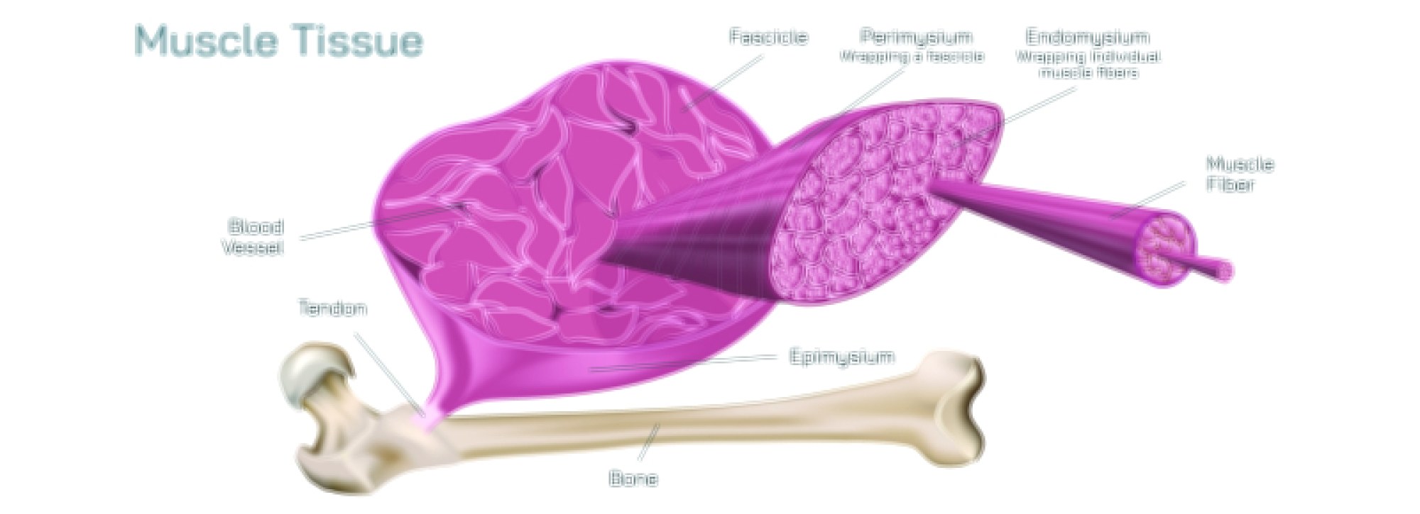
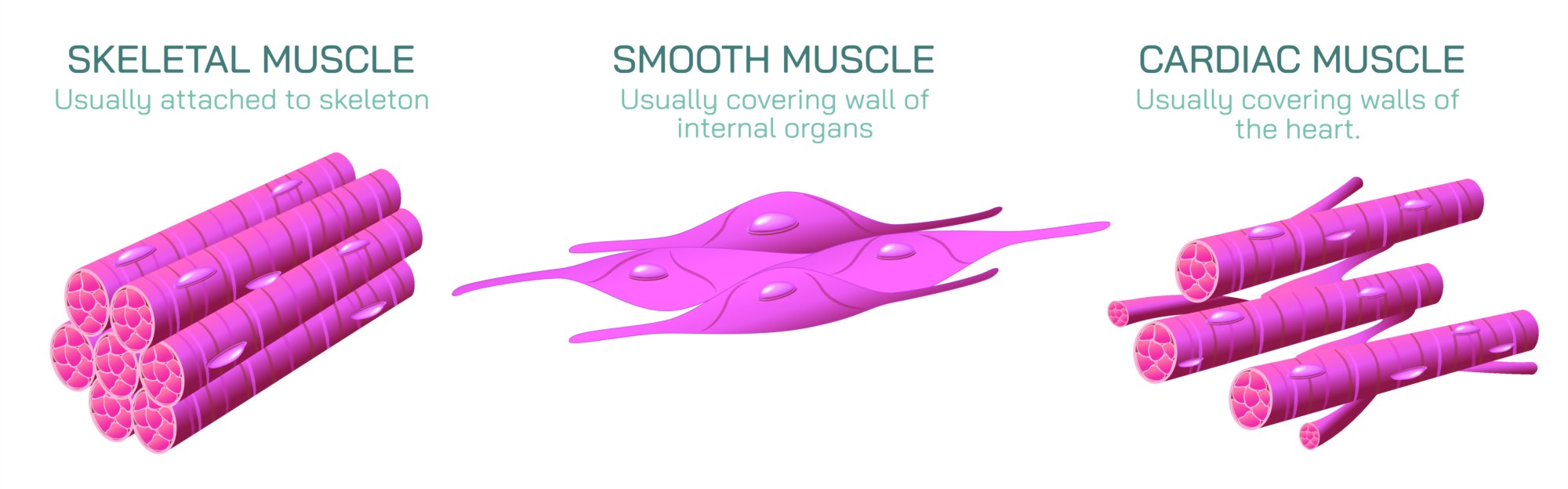
Skeletal muscle is the most familiar form, anchored to bones through tendons and responsible for locomotion, speech, facial expression, and nearly every voluntary movement. Under the microscope, skeletal muscle fibers reveal an extraordinary level of structural organization, appearing as long cylindrical multinucleated cells arranged in parallel bundles. Their most distinctive feature is the presence of striations, the alternating light and dark bands formed by the precise alignment of actin and myosin filaments within repeating contractile units called sarcomeres. These sarcomeres make skeletal muscle capable of fast, forceful, and highly controlled contractions. Each fiber is wrapped in connective tissue, and groups of fibers form fascicles, which together create whole muscles. Motor neurons extend from the spinal cord and brain to skeletal muscle fibers, where they form neuromuscular junctions that release acetylcholine, triggering contraction. This direct neural control allows for fine voluntary adjustments of force, enabling activities ranging from lifting heavy weights to the delicate motion required to write or play a musical instrument. The energy demands of skeletal muscle are immense and are supported by abundant mitochondria, creatine phosphate stores, and highly dynamic ATP production that allows quick response to suddenly changing workloads.
Smooth muscle differs radically in appearance and function. It does not form long multinucleated fibers, nor does it contain visible striations, instead presenting as short spindle-shaped cells with a single nucleus. Although smooth muscle contains actin and myosin, these filaments are arranged in a mesh-like pattern rather than in organized sarcomeres, allowing contraction in multiple directions. Smooth muscle lines the internal organs and passageways of the body—within the stomach, intestines, bladder, uterus, blood vessels, and respiratory system—where its slow, sustained, and rhythmic contractions maintain essential bodily processes that do not require conscious thought. Rather than direct voluntary innervation, smooth muscle is controlled by the autonomic nervous system, hormones, and local chemical signals. The gastrointestinal tract uses smooth muscle contractions in waves called peristalsis to move food; blood vessels constrict or relax through smooth muscle to regulate blood pressure and flow; and the uterus relies on powerful smooth muscle activation during childbirth. Because smooth muscle can maintain tension for long periods without fatigue and with minimal energy expenditure, it functions continuously throughout life and adjusts quietly to the internal demands of digestion, circulation, and respiration.
Cardiac muscle stands as the most specialized muscle type, built exclusively for the continuous contraction required to pump blood throughout the entire lifespan. Like skeletal muscle, cardiac muscle contains striations because actin and myosin are organized into sarcomeres; however, its fibers are branched, shorter, and connected end-to-end through intercalated discs, which appear as dark junctional bands under magnification. These discs contain gap junctions and desmosomes that allow electrical impulses to pass rapidly between cells and maintain structural integrity during contraction. This arrangement turns the myocardium into a fully coordinated functional syncytium, enabling the heart to contract as a single unit rather than as independent fibers. Unlike skeletal muscle, cardiac muscle does not require conscious control, and unlike smooth muscle, it follows a rhythmic pattern with extraordinary precision. Cardiac cells contain specialized pacemaker regions, most notably the sinoatrial node, which generates electrical impulses autonomously. These impulses spread through the conduction system of the heart, triggering timed contraction of the atria and ventricles with a pattern stable enough to maintain an uninterrupted heartbeat for decades. Cardiac muscle has an exceptionally high density of mitochondria, reflecting its need for constant ATP generation to sustain uninterrupted activation. Its reliance on oxygen-rich blood is so great that even brief deprivation can lead to irreversible cell damage.
Although skeletal, smooth, and cardiac muscles are structurally and functionally distinct, they also demonstrate continuous cooperation. A person running involves skeletal muscle powering the stride, smooth muscle regulating blood flow and directing oxygen to working tissues, and cardiac muscle increasing heart rate and contraction strength to meet metabolic demands. The body’s survival rests on this seamless integration. Skeletal muscle allows purposeful interaction with the world, smooth muscle regulates the internal environment, and cardiac muscle sustains the circulation that links all cells of the body. What unites the three muscle types is their dependence on the sliding filament mechanism, yet what differentiates them is the degree of voluntary control, the speed and pattern of contraction, and the organizational design of their cellular machinery. By recognizing how these muscles complement one another rather than simply operate independently, it becomes clear that human movement and life itself emerge from a biological system built on specialization, coordination, and robust adaptability.