MRI Scan Gradient Echo: Imaging Technique and Signal Formation
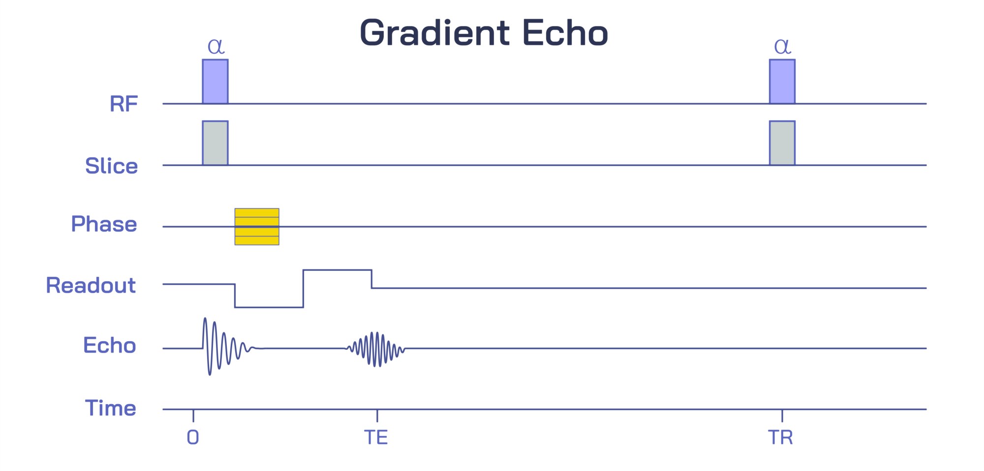
Magnetic Resonance Imaging relies on the manipulation and detection of hydrogen protons inside the body, but gradient echo imaging represents one of the most unique and versatile ways of transforming proton behavior into a clinically useful signal. In this form of MRI, the key idea is that the echo is not produced by a refocusing radiofrequency pulse, but instead by the careful application of magnetic field gradients that dephase and then rephase spin coherence in the transverse plane. To understand this technique in a continuous narrative, it helps to imagine the journey of magnetization from the instant the hydrogen protons align with the primary magnetic field to the moment a measurable echo forms. In a strong magnetic field, billions of protons align their magnetic vectors and precess at a frequency determined by tissue environment. Gradient echo imaging begins when a radiofrequency excitation pulse tips the net magnetization from the longitudinal axis toward the transverse plane. Unlike the 90-degree pulse used in spin-echo sequences, gradient echo imaging often uses a low flip-angle pulse, creating a partial rotation of magnetization that leaves substantial longitudinal component available for rapid repetition. This is the first reason gradient echo imaging can acquire images far more quickly than conventional spin echo: magnetization recovery is faster because it is never fully saturated.
As soon as the excitation pulse ends, the transverse magnetization begins to decay due to phase differences between protons. Some of these differences arise from interactions between neighboring spins, and some arise from local disturbances in the magnetic field. In spin echo sequences, a 180-degree pulse flips these out-of-sync spins so they rephase later. Gradient echo imaging does not use such a refocusing pulse; instead, it allows dephasing to occur naturally and then actively reverses it using magnetic field gradients. When the frequency-encoding gradient or a specially applied gradient lobe first exaggerates the loss of phase coherence and a following gradient of opposite polarity restores synchronization, the proton ensemble regains alignment momentarily. At the instant when the spins once again move together in phase, the signal peaks and forms what is called the gradient echo. This rephasing, however, never fully compensates for all sources of dephasing, because it corrects only for spatially induced frequency offsets and not intrinsic spin-spin interactions. Therefore, the signal decays not according to the T2 relaxation constant but a faster effective constant known as T2*. This subtle distinction defines the contrast and sensitivity of gradient echo images.
Signal Formation and Contrast in Gradient Echo Imaging
Because gradient echo imaging depends heavily on T2* rather than T2, it becomes exquisitely sensitive to microscopic magnetic field variations caused by iron, calcium, deoxygenated blood, and metallic implants. When the echo time, the interval between the excitation pulse and the echo peak, is relatively long, susceptibility effects have more time to act and signal loss becomes prominent near deposits of hemosiderin, microbleeds, or venous blood containing deoxyhemoglobin. This turns gradient echo into a powerful tool for detecting hemorrhage, trauma, neurodegeneration, and vascular abnormalities. Conversely, the echo time can be kept extremely short to reduce these effects and instead emphasize differences in T1 relaxation or proton density, making gradient echo adaptable across widely differing diagnostic aims. Another crucial parameter is the flip angle. With a short repetition time, a low flip angle allows steady-state equilibrium of longitudinal magnetization to develop, tailoring the image contrast to highlight fat, fluid, dynamic contrast transport, or moving structures such as the beating heart. Because the sequence never fully erases transverse magnetization unless purposely spoiled, the contrast behavior changes dramatically based on whether the sequence is designed to dephase residual signal or preserve it.
“Spoiled” gradient echo sequences intentionally eliminate leftover transverse coherence using gradient spoiling or radiofrequency spoiling. This forces the magnetization to behave in a predictable manner controlled by T1 weighting. In enhanced imaging after contrast injection, spoiled gradient echo is ideal because the high T1 sensitivity highlights gadolinium-enhanced tissues vividly. At the other end of the contrast spectrum lies the balanced steady-state free precession approach. This variation preserves both transverse and longitudinal components, stabilizing the signal into a high-intensity steady state. Fluids such as blood and cerebrospinal fluid appear bright in these images, making balanced sequences invaluable in cardiac cine imaging and neuroanatomy. Between these two extremes sits the susceptibility-weighted domain, which applies long echo times on purpose to capture subtle dephasing effects. This application of gradient echo has transformed brain imaging, making it possible to detect minute microhemorrhages, venous structures, iron deposits, and traumatic injuries invisible in other MRI sequences.
Clinical and Technical Advantages of Gradient Echo MRI
The advantages of gradient echo imaging begin with speed. Because there is no 180-degree refocusing pulse, repetition times can be extremely short and energy deposition remains low. This allows rapid three-dimensional acquisitions of whole anatomical regions with thin slices and high resolution, enabling visualization of complex structures such as cartilage, brain cortex, heart chambers, pulmonary vasculature, and abdominal organs in unprecedented detail. Real-time imaging of motion becomes feasible, allowing physicians to observe cardiac contraction, respiratory motion, and contrast flow through blood vessels dynamically. In functional MRI, gradient echo is central to blood oxygen level–dependent imaging. The method harnesses the fact that deoxygenated hemoglobin creates microscopic field distortions that affect T2* decay. Because neural activation changes the oxygenation of local blood flow, small fluctuations in gradient echo signal can be decoded into maps of brain activity. Thus, gradient echo sequences expand beyond morphology into physiology, metabolism, circulation, and neurocognitive research.
However, the strength of gradient echo can also become a limitation. Because susceptibility effects are amplified rather than suppressed, the presence of surgical implants, dental fillings, orthopedic metalwork, and air–tissue interfaces can cause signal dropout and distortion. Gradient echo sequences require thoughtful tuning of parameters to minimize artifacts while preserving diagnostic information. This trade-off between sensitivity and artifact is why multiple variations of gradient echo were developed, each one balancing speed, contrast, resolution, and artifact control differently. The entire family of sequences—including spoiled GRE, ultrashort echo GRE, balanced SSFP, susceptibility-weighted imaging, and time-resolved contrast-enhanced angiography—illustrates that gradient echo imaging is not a single method but an adaptable framework that can be reshaped to match clinical needs.
In its complete conceptual form, gradient echo imaging traces a circular cycle of magnetization behavior: spins are partially tipped, allowed to dephase, driven back into phase by gradients, used to generate a detectable signal, and then rapidly excited again. Every part of the sequence builds on the natural physics of hydrogen protons rather than overpowering them. This efficiency enables rapid image acquisition, adjustable tissue contrast, sensitivity to biological processes, and compatibility with complex three-dimensional and time-resolved imaging. The gradient echo technique makes MRI faster without sacrificing depth, more physiologically revealing without abandoning structural clarity, and more flexible without losing diagnostic precision. It demonstrates that medical imaging can evolve not by forcing nature into compliance, but by harnessing the inherent patterns of magnetization and transforming them into visual information that reveals both anatomy and function within the human body.