Thyroid Anatomy – Structure, Function, Hormone Regulation, Vascular Network, Cellular Composition, and its Essential Role in Human Metabolism and Endocrine Health
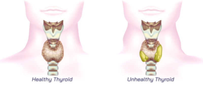
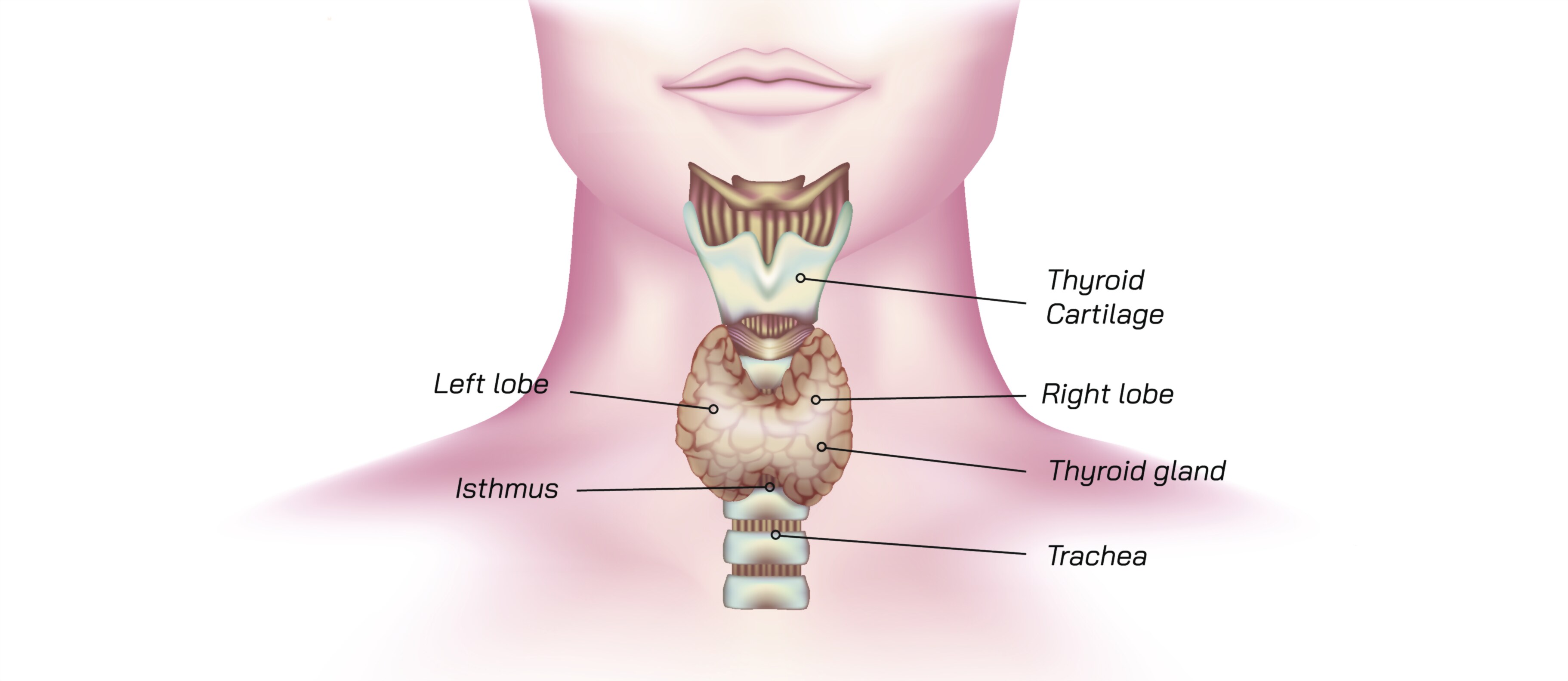
The thyroid gland is one of the most important organs of the human endocrine system because it controls the body’s metabolic rate, energy balance, temperature regulation, and growth through the production of thyroid hormones. Although small in size, the thyroid influences nearly every cell and organ in the body, making its anatomy and structure vital to understanding overall human physiology. Located at the base of the neck, just below the larynx and in front of the trachea, the thyroid has a shape commonly compared to a butterfly, with two lateral lobes—right and left—connected by a narrow bridge called the isthmus. The gland wraps partially around the trachea like a protective shield, which is why the ancient Greek word “thyreos,” meaning shield, contributed to its name. Its position in the neck allows strong vascular supply and efficient hormonal release into the bloodstream, supporting the gland’s fast and constant regulatory activity.
Structural Organization and Lobes of the Thyroid
The thyroid’s external form is smooth and soft, and although its average weight ranges from 15 to 25 grams, it can vary significantly in size across individuals based on age, gender, iodine intake, and physiological conditions such as puberty or pregnancy. Each lobe of the thyroid extends upward toward the thyroid cartilage and downward along the trachea, with the isthmus laying across the second and third tracheal rings. In some people, a small lobe called the pyramidal lobe extends upward from the isthmus, a remnant of embryological development showing where the gland originally migrated from the back of the tongue. This developmental history reminds students that endocrine anatomy is closely tied to embryology, where migration of tissues determines their adult placement and relationships with surrounding structures.
Microscopic Structure and Hormone-Secreting Cells
Beneath its smooth surface, the thyroid has a highly organized microscopic architecture. The gland is composed of countless spherical units called follicles, which form the basic functional and structural components of thyroid tissue. Each follicle consists of a central cavity filled with a jelly-like material called colloid—a dense storage form of thyroid hormones—and a surrounding layer of epithelial cells known as follicular cells. These follicular cells synthesize and secrete the primary thyroid hormones, thyroxine (T4) and triiodothyronine (T3), by absorbing iodine from the bloodstream and attaching it to tyrosine molecules within the colloid. When the body signals a need for more thyroid hormone, follicular cells retrieve colloid material and release T3 and T4 into the blood.
Inside the thyroid are also parafollicular cells, or C cells, which lie between follicles and secrete the hormone calcitonin. Calcitonin helps regulate calcium levels by lowering calcium concentration in the blood when it becomes too high, complementing the actions of the parathyroid glands. Although calcitonin plays a smaller role than other calcium-regulating hormones in humans, the presence of C cells shows that the thyroid contributes not only to metabolic control but also to mineral balance.
Hormonal Function and Endocrine Regulation
4. Once levels rise in the bloodstream, the hormones circulate throughout the body and bind to receptors in nearly all tissues—not to trigger immediate actions like neurotransmitters, but to regulate gene expression over time. T3 and T4 increase the metabolic rate, enhance oxygen consumption, regulate ATP production, influence heart rate, help maintain body temperature, and play major roles in fetal brain development and childhood growth. When T3 and T4 levels become adequate, they send negative feedback to the pituitary and hypothalamus to reduce TSH and TRH output, maintaining homeostasis. This negative feedback loop ensures that thyroid function remains balanced and prevents overproduction or underproduction of hormones.
Blood Supply, Innervation, and Lymphatic Drainage
The thyroid’s intense metabolic activity demands a rich blood supply. The gland receives arterial blood primarily from the superior thyroid arteries, originating from the external carotid arteries, and the inferior thyroid arteries, arising from the thyrocervical trunks of the subclavian arteries. In some individuals, an additional artery known as the thyroid ima artery may also supply the gland. Venous drainage occurs through a network of superior, middle, and inferior thyroid veins, which carry blood from the gland back toward the heart. This dense network allows rapid endocrine diffusion and also contributes to the tendency for heavy bleeding during thyroid surgery if proper vascular control is not maintained. Innervation arises from sympathetic fibers that modulate blood flow rather than directly stimulating hormone release, reflecting that thyroid hormone production is governed by the endocrine system rather than direct neural commands. Lymph vessels from the thyroid drain into cervical lymph nodes, which is medically relevant because certain thyroid diseases can involve lymphatic spread.
Relationship With Nearby Structures and Clinical Relevance
Because of its location, the thyroid sits close to several important anatomical structures. The recurrent laryngeal nerves run behind the thyroid and innervate the vocal cords, which is why thyroid enlargement or surgery may affect speech when these nerves are involved. The parathyroid glands, located on the posterior surface of the thyroid, regulate calcium homeostasis, so surgical procedures require precision to preserve them. Goiter, thyroid enlargement, may compress the trachea or esophagus, causing breathing or swallowing difficulties, showing how changes in gland size can impact surrounding anatomy. Although the thyroid is small, disorders such as hypothyroidism, hyperthyroidism, thyroid nodules, autoimmune disease, and thyroid cancer can produce wide-ranging body effects because hormone imbalance disrupts metabolism at a systemic level.
The Thyroid as a Model of the Connection Between Anatomy and Whole-Body Physiology
Studying thyroid anatomy helps learners understand that the structure of an organ determines its function and that even microscopic changes in hormone production can alter metabolism, temperature, digestive speed, cardiovascular function, growth, fertility, and neurological development. It also demonstrates how endocrine organs act with precision rather than through constant activity; the thyroid produces hormones slowly and releases them as needed, integrating with brain regulation and feedback systems. The gland illustrates how nutrition affects physiology because adequate iodine intake is critical for hormone synthesis. It also reveals the delicate balance of the endocrine system, showing that small imbalances in regulation can have profound consequences for energy, mood, cognition, and physical development.
Ultimately, the thyroid offers a powerful educational example of how biology connects microstructure to macroscale health. Its butterfly-shaped anatomy, follicular architecture, hormonal output, vascular supply, and regulatory relationships form a compact yet immensely influential organ that sustains metabolism across the lifespan. Through the study of thyroid anatomy, students gain not only a foundational understanding of endocrine physiology but also a deeper appreciation for how finely tuned systems within the human body maintain equilibrium and support life.