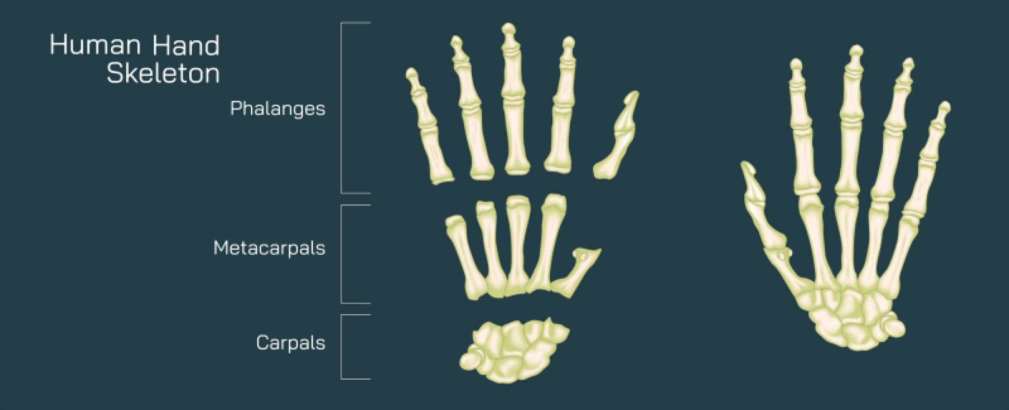
Human Eye Anatomy Illustration: Detailed Structure of Eye and Its Functional Parts
The human eye is one of the most sophisticated biological optical systems, and an illustration that breaks down its anatomy allows us to appreciate not only the complexity of its structure but also the remarkable way in which its parts work together to transform light into vision. When viewed in cross-section—typically through a longitudinal cut running from front to back—the eye reveals a layered and compartmentalized design optimized to capture light, focus it precisely, detect it through specialized photoreceptors, and send the resulting information to the brain. Although the eye is small, roughly spherical, and only about 2.5 centimeters in diameter, every region performs a precise role in the sequence that creates sight. A detailed anatomical diagram of the eye highlights this seamless flow: light enters the eye, bends (refracts), focuses onto the retina, and ultimately becomes electrical signals interpreted by the brain as images.
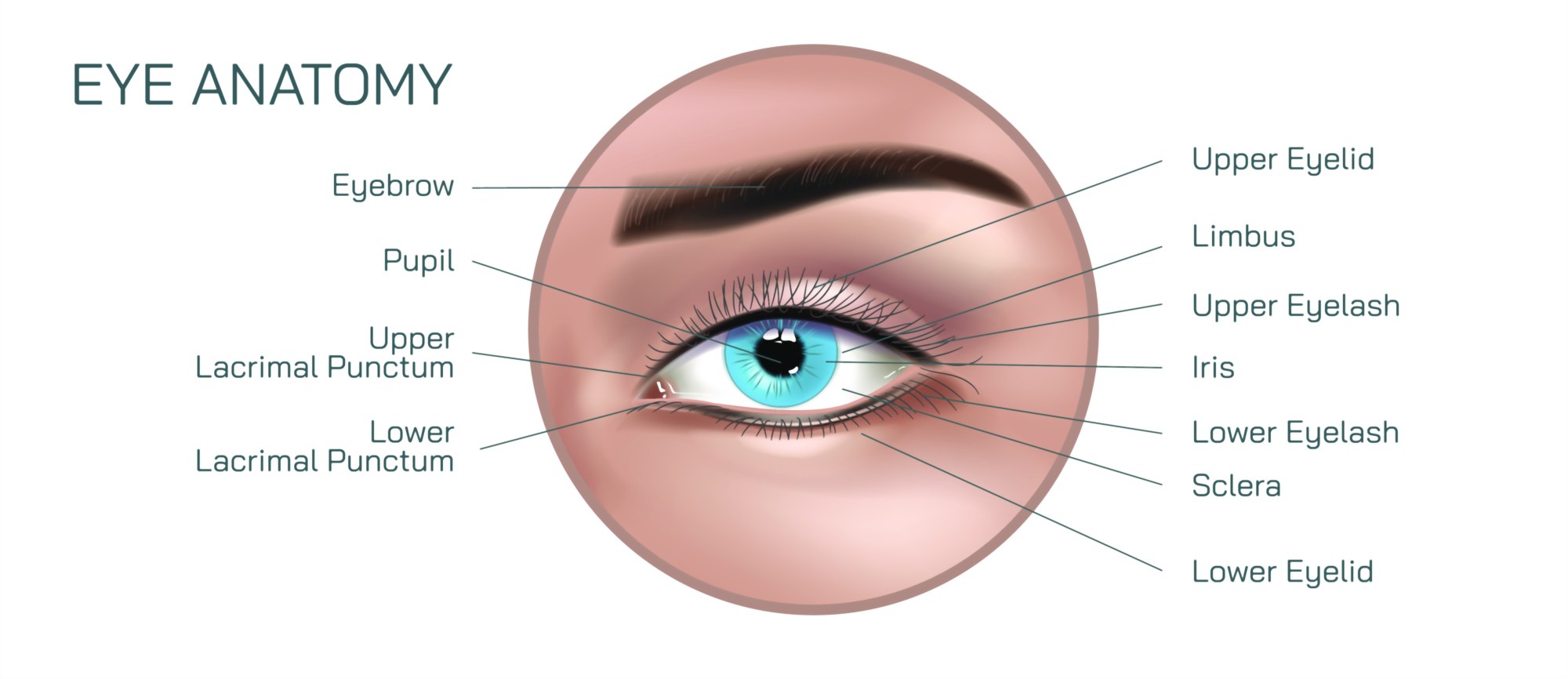
At the outermost level of the eye illustration, the viewer encounters the cornea, the transparent, curved front window of the eye. This structure performs the majority of the eye’s focusing power because its rounded surface sharply bends incoming light toward the interior. In diagrams, the cornea usually appears as a bulging clear layer overlaying the front of the eye and is shown continuous with the sclera, the tough, opaque white coat that encases the rest of the eyeball and maintains shape and protection. Together, the cornea and sclera form the fibrous tunic, the outermost layer of the eye. The junction where the clear cornea meets the white sclera—known as the limbus—is often labeled to show where the tissue transitions and where fluid drainage channels used in glaucoma regulation are located.
Behind the cornea lies the anterior chamber, filled with aqueous humor, a clear fluid that maintains intraocular pressure and nourishes the cornea and lens. This fluid flows continuously and drains through a microscopic network called the trabecular meshwork, illustrated near the limbus. A disruption of this flow can increase pressure and damage the optic nerve, a condition depicted in glaucoma diagrams. Surrounding this chamber is the colored iris, the circular muscle that regulates the amount of light entering the eye. The iris illustration almost always features its central opening, the pupil, which widens (dilates) or narrows (constricts) automatically in response to light intensity. In bright daylight, the iris constricts the pupil to protect sensitive photoreceptors; in dim light, it relaxes to enlarge the pupil and allow more light to enter.
Directly behind the pupil sits the lens, a transparent, flexible, biconvex structure responsible for fine-tuning focus. The lens changes shape through the action of the surrounding ciliary body and its suspensory ligaments (zonular fibers). When the ciliary muscles contract, the lens becomes more rounded, allowing the eye to focus on near objects. When the muscles relax, the lens flattens for distant focus. Illustrations that depict these actions show arrows compressing or stretching the lens depending on the visual state. This focusing mechanism, called accommodation, weakens with age as the lens stiffens, a condition known as presbyopia—a feature sometimes shown in age-progressive diagrams.
Behind the lens lies the largest chamber of the eye, filled with vitreous humor, a clear gel that maintains the spherical shape of the globe and keeps the retina pressed smoothly against the back wall. Floating debris within this gel—sometimes shown as fibers or dots—cause “floaters,” which appear in vision because the gel casts shadows on the retina. The interior rear surface of the eye contains the retina, the paper-thin yet highly specialized neural tissue where light is converted to nerve signals. Illustrations highlight the retina as a multilayered sheet containing rods and cones, the photoreceptors responsible for detecting light and color. Rods support night and peripheral vision, highly sensitive to low light but not color; cones enable sharp, detailed, color vision and cluster densely in the fovea, a small depression in the retina that represents the point of greatest visual acuity.
The fovea sits at the center of the macula, a yellowish area responsible for precise central vision. In many anatomical diagrams, the macula and fovea are emphasized to show why degenerative conditions affecting this area—like macular degeneration—impair reading, facial recognition, and fine detail work. Surrounding the retina is the choroid, a dark, vascular layer that delivers oxygen and nutrients to the retina while absorbing stray light to prevent internal reflections that would blur vision. Just outside the choroid lies the sclera, completing the layered structure like concentric shells.
From the surface of the retina emerges the optic nerve, composed of over a million nerve fibers carrying visual information toward the brain. Where the optic nerve exits the eye lies the optic disc, also called the blind spot, because this region contains no photoreceptors. In illustrations, the optic disc appears as a pale round area where blood vessels converge before leaving the eye. Although the blind spot exists anatomically, the brain fills in missing information through contextual interpretation, making it unnoticeable during everyday vision. The trajectory of the optic nerve shown in diagrams soon leads to the optic chiasm, where some nerve fibers cross, enabling binocular vision and depth perception through visual field integration from both eyes.
In addition to mapping structural pathways, many eye illustrations highlight the protective and supportive systems surrounding the eyeball. The conjunctiva lines the inside of the eyelids and covers the sclera as a thin, transparent membrane that helps lubricate and protect the eye from foreign particles. The lacrimal gland, located above the outer corner of the eye, produces tears that wash across the corneal surface before draining through the nasolacrimal duct into the nasal cavity. Muscles controlling eye movement—called the extraocular muscles—are often shown attached to the sclera, enabling precise rotation of the eyeball. These include the superior, inferior, lateral, and medial rectus muscles, along with the superior and inferior oblique muscles. Illustrations of these muscles demonstrate how rapid, coordinated movements create smooth tracking, depth perception, and stabilization of vision during head motion.
A detailed diagram of the cornea, lens, and retina working together reinforces the functional process of refraction and image formation. Light rays shown entering the cornea bend sharply, pass through the aqueous humor, iris opening, and lens, and then converge precisely on the fovea. If light converges in front of the retina, the illustration represents myopia (nearsightedness); if light converges behind the retina, it represents hyperopia (farsightedness). In these visual comparisons, corrective lenses are drawn to redirect light appropriately. Laser eye surgery diagrams show reshaping of the cornea to correct refractive errors by altering how strongly it bends light.
The retina itself—though often depicted as a single region—is a remarkably complex neural processing hub. Magnified illustrations show rod and cone photoreceptors synapsing with bipolar and ganglion cells, forming pathways that integrate visual signals before they ever reach the brain. This organization allows contrast detection, motion sensitivity, and preliminary image sharpening to happen directly in the eye rather than relying exclusively on the brain. Following this, electrical impulses travel via the optic nerve to the visual cortex in the occipital lobe, where the brain constructs perception from the signals.
Ultimately, a human eye anatomy illustration goes beyond anatomical labeling—it narrates the complete biological process of vision from the first moment light enters the eye to the point the brain interprets the message. It reveals how delicate tissues and precisely arranged structures function in harmony to provide clarity, color, depth, and motion perception. It shows why the eye is not just a camera-like device but a living extension of the nervous system, constantly adjusting and processing in real time. Through a combination of layered anatomy, fluid circulation, optical physics, and neural transmission, the illustration reveals that the eye is not merely a sensory organ—it is the first stage of vision itself, a biological masterpiece linking the external world to human consciousness.