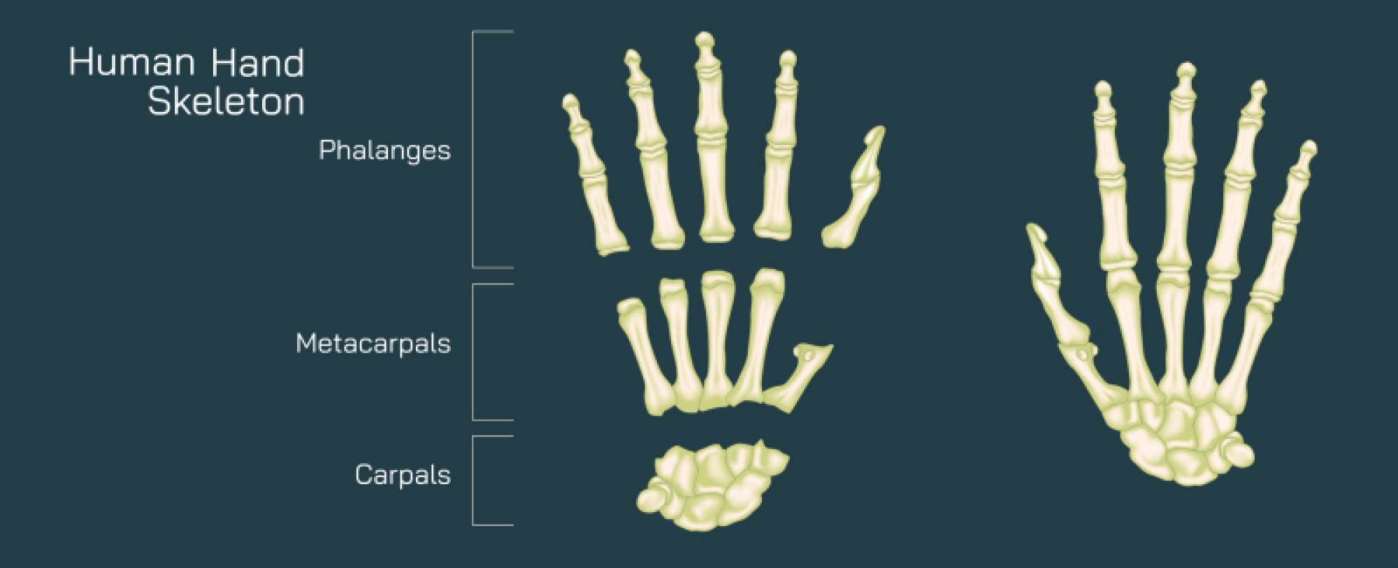
Body Fat Percentage Illustration: Healthy Ranges and Fat Distribution in the Human Body
Body fat percentage is one of the most informative measurements for evaluating physical health because it describes not just weight, but the composition of the human body. Unlike basic weight readings, which cannot distinguish muscle from fat or bone from water, body fat percentage reveals how much of a person’s body mass is composed of fat tissue. When illustrated visually, this concept becomes far more meaningful, because diagrams show the difference between lean body mass and stored fat in various regions of the body, painting a clear picture of how fat distribution influences shape, health, metabolism, and disease risk. A realistic illustration of body fat percentage does not label the body as simply “thin” or “overweight,” but instead depicts the complex variation of fat layers beneath the skin and around internal organs. Fat is not merely a passive storage depot; it is an active tissue involved in hormone regulation, energy balance, insulation, and protection. Its amount and distribution can support health when within balanced ranges or strain the body when excessively accumulated or misplaced.
Healthy ranges of body fat vary with age, sex, and biological demands, something clearly highlighted in educational illustrations that differentiate typical profiles of men and women. In general, men have naturally lower body fat percentages because of their hormonal environment and greater lean muscle mass, whereas women carry more fat due to reproductive biology and estrogen-driven fat storage in the hips, thighs, and chest. A diagram of healthy ranges often shows men in the approximate range of 10–20% and women in the approximate range of 18–28% during adulthood, with ranges increasing gradually with age due to metabolic changes. These healthy levels represent the amount of fat needed to sustain energy reserves, regulate body temperature, cushion internal organs, and maintain hormonal balance. In contrast, essential fat—shown in some diagrams as the minimum biological requirement—exists at even lower ranges (around 3–5% in men and 8–12% in women) and is necessary for survival because it supports critical cell structures, nerve insulation, and reproductive function. When diagrams compare essential body fat, healthy ranges, athletic ranges, and excessive fat categories, viewers can visually understand that fat is not inherently negative; it becomes harmful only when its quantity or distribution exceeds what the body can handle safely.
To illustrate body fat distribution visually, diagrams often highlight two primary types of fat storage: subcutaneous fat and visceral fat. Subcutaneous fat sits directly beneath the skin and is responsible for the softness or curvature of the body surface. It is commonly located in the thighs, hips, lower abdomen, arms, and buttocks. In moderation, it serves as insulation and helps protect against physical impact. When excessive, it becomes the source of aesthetic changes that are visible externally, such as thigh thickness or abdominal fullness. Visceral fat, however, lies hidden deeper in the body around the internal organs of the abdomen. It does not significantly alter outward appearance in early stages and can exist even in individuals who appear slim. Illustration cross-sections often highlight visceral fat as a thick yellow ring surrounding organs such as the liver, stomach, and intestines. Visceral fat is metabolically active, releasing inflammatory chemicals and hormone-like signals that can disrupt glucose metabolism, blood pressure, and lipid levels. For this reason, diagrams clearly differentiate the two, demonstrating that fat located on the surface of the body is not always as medically dangerous as fat accumulating internally near vital organs.
Different categories of fat distribution produce recognizable body shapes when translated into illustration form. Body fat that accumulates around the hips and thighs is often described as a “pear-shaped” pattern and is more common in biological females due to estrogen-mediated fat storage. Although large in volume, this distribution tends to pose fewer cardiovascular risks because it represents primarily subcutaneous fat stores. Body fat that accumulates around the abdomen and waist creates an “apple-shaped” pattern, more frequently associated with males but seen in females after menopause or with hormonal imbalance. This region is associated with higher visceral fat storage, and illustrated diagrams show that this pattern corresponds with elevated health risks such as diabetes, fatty liver disease, heart disease, and metabolic syndrome. By comparing both patterns side by side, illustrations help viewers understand that where fat is stored matters just as much as how much of it is present. Fat distributed around the stomach and midsection is far more concerning than fat concentrated in the lower body simply because of its relationship with internal organ stress.
Body fat percentage changes dramatically across different lifestyles, activity levels, and aging processes, and diagrams reflecting these timelines help people understand how fat distribution shifts naturally across decades. Young adults tend to display a balanced distribution with moderate subcutaneous fat and relatively low visceral fat. As individuals age, metabolism slows, hormone levels decline, and muscle mass gradually diminishes unless physically maintained. Illustrations depicting aging show that fat begins to accumulate more around the abdomen, while depots in the limbs decrease. This trend applies to both men and women, though its timing and extent vary individually. Illustrations that plot body fat percentage changes over time reinforce that fat accumulation is not merely a function of diet but also of biological aging and muscle loss. This is why diagrams of body fat percentage often include the concept of lean mass preservation, emphasizing that maintaining muscle through resistance training, daily movement, and protein intake helps stabilize metabolism and slow fat gain.
Another important visual theme that consistently appears in body fat illustrations is the distinction between healthy and unhealthy low fat levels. Many people assume that the lowest possible body fat is always ideal, but the visual comparison of different ranges demonstrates otherwise. When a diagram shows an extremely low-fat body, often associated with competitive bodybuilding or severe calorie restriction, it displays a loss of cushioning between organs, hormonal disruption, reduced immune performance, and impaired reproductive function. Muscles and veins may appear extremely defined, but the biological stress is immense. Thus, the healthiest physical state lies not at the far ends of the fat percentage spectrum but in the middle ranges, where the body is fully functional, hormonally stable, and metabolically supported. Illustrations that contrast “athletic lean,” “healthy natural,” and “extremely low fat” bodies help people see beauty and biological well-being not as contradictions but as balance points.
In addition to external diagrams, some educational illustrations zoom inside the metabolic system to show how fat cells function. Under the microscope, fat cells—or adipocytes—appear as round droplets storing triglycerides. When body fat percentage increases, these cells expand like balloons rather than multiplying rapidly; only after significant storage do new fat cells develop. When the percentage decreases, these cells shrink but rarely disappear completely. This cellular illustration communicates why long-term weight management requires lifestyle changes rather than temporary dieting—once fat cells exist, they remain metabolically prepared to store fat again. Some diagrams extend even further to show how fat cells release hormones such as leptin, which influences hunger and energy regulation. This internal view reinforces that fat is biologically active and deeply integrated into body systems.
Ultimately, a body fat percentage illustration is about understanding—not judgment. It shows the broad spectrum of human body composition and teaches that health depends on multiple factors: the volume of fat, the distribution of fat, the stability of metabolism, and the balance between muscle and fat over time. It visualizes fat as an essential component of the body rather than a flaw to be removed. Learning through these comparisons encourages individuals to shift focus from scale weight and appearance alone to deeper measures of health, such as the resilience of the cardiovascular system, the condition of the metabolic organs, the balance of hormones, and the long-term sustainment of muscle strength. When people understand body fat in this way, they gain a more compassionate and scientific perspective of the human form—one that celebrates the body as a biological system always working to preserve life, stability, and equilibrium.