Melanin Pigments and Skin Tone Types Illustration: Eumelanin, Pheomelanin, and Skin Color Variations Explained
Human skin color is shaped by one of the most fascinating biological pigments on Earth: melanin. An illustration comparing eumelanin, pheomelanin, and how their distribution influences skin tone variations provides a compelling visual understanding of why humans display such a wide spectrum of skin shades worldwide. Skin color is not determined by the number of melanocytes alone—because nearly all humans have approximately the same number of these pigment-producing cells. Instead, diagrams reveal that differences emerge from how much melanin is produced, the type of melanin synthesized, how pigments are packaged and transported, and how deeply they are deposited within the skin.
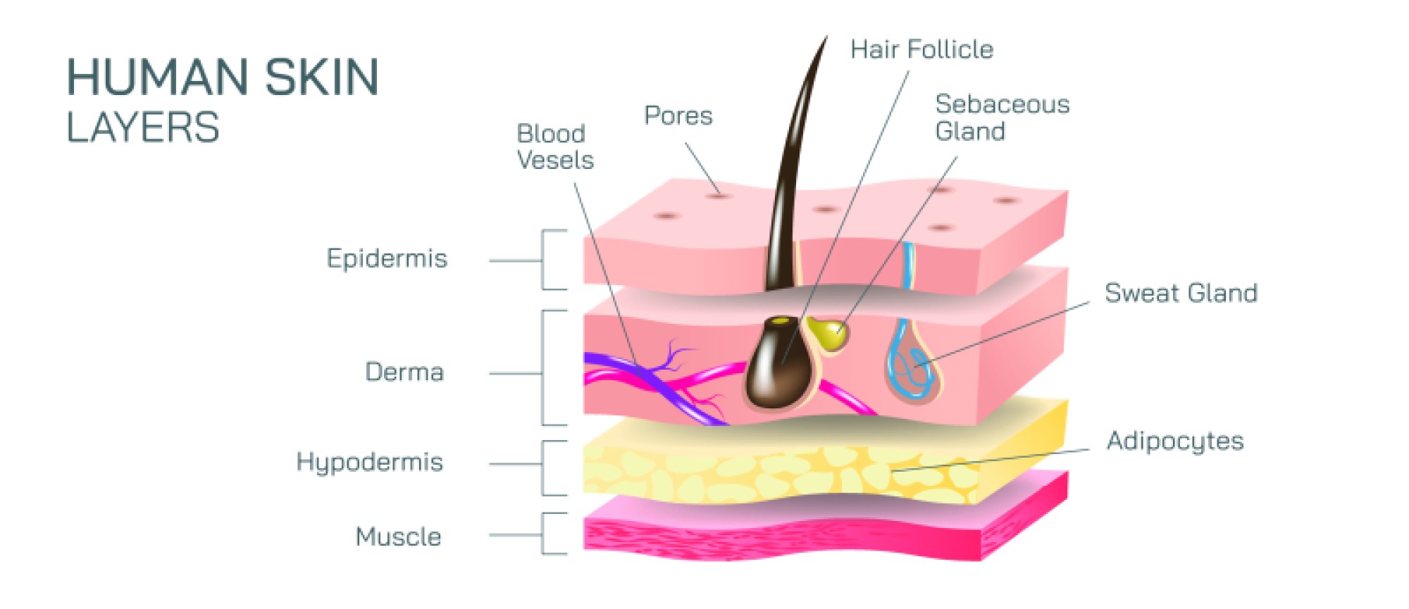
A well-structured illustration begins with the location of melanin production. Zoomed-in diagrams of the epidermis show melanocytes residing in the basal layer, interspersed among keratinocytes. Each melanocyte contains specialized organelles called melanosomes, where melanin pigment is synthesized. These melanosomes are then transferred to surrounding keratinocytes through microscopic dendritic extensions, distributing pigment across the skin. In illustrations, melanocytes are often drawn as star-shaped cells with branching arms, delivering melanin-filled vesicles upward throughout the epidermis. The scattering of these pigmented cells visually reinforces that skin color depends not on melanocyte quantity, but on the size, number, and dispersion pattern of melanosomes.
From here, illustrations typically branch into the two main types of melanin:
Eumelanin
Eumelanin is the brown-black pigment responsible for most of the dark coloration in human hair, skin, and eyes. Molecular diagrams depict eumelanin as a dense, irregular polymer structure capable of absorbing large amounts of ultraviolet (UV) radiation. Biologically, this pigment offers powerful protection against sunlight by neutralizing free radicals and preventing UV-induced DNA damage. In visual comparisons, high-eumelanin skin types are shown with large, numerous melanosomes that are heavily pigmented and dispersed individually throughout keratinocytes. This pattern preserves pigment longer as cells migrate toward the skin surface, resulting in darker skin tones and greater natural UV resistance.
Pheomelanin
Pheomelanin is the yellow-red pigment found prominently in red hair, lighter skin, freckles, and lips. Structural illustrations show pheomelanin with sulfur-containing molecules—in contrast to the polymer makeup of eumelanin. Unlike eumelanin, pheomelanin absorbs less UV radiation and offers weaker shielding from sunlight. In skin tone diagrams, pheomelanin appears in smaller, lighter melanosomes that tend to cluster together rather than disperse individually. This clustering contributes to lighter skin tones and increased UV sensitivity, especially in individuals with high pheomelanin production paired with low eumelanin levels.
Blended Pigmentation and Skin Tone Variation
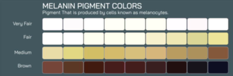
A key feature of melanin illustrations is the gradient or spectrum of skin color across populations, from very light to very dark. This gradient is not caused by a binary switch between eumelanin and pheomelanin, but by varying ratios of the two pigments combined with differences in the amount of melanin synthesized and retained within the epidermis. For example:
Very light skin tones may be illustrated with low total melanin and a higher proportion of pheomelanin.
Olive and medium tones display moderate melanin levels with a mixture of eumelanin and pheomelanin.
Deep brown and black skin tones show high levels of eumelanin and dense epidermal melanosome distribution.
Some illustrations also depict skin reflectance curves, demonstrating how melanin strongly absorbs UV and visible wavelengths and drastically reduces reflectivity as eumelanin concentration increases.
Melanin and UV Radiation
Side-by-side UV exposure diagrams highlight how melanin protects DNA from UV damage. In darker skin illustrations, eumelanin forms an efficient UV shield by absorbing and scattering radiation before it reaches deeper tissues. In lighter skin illustrations, higher UV penetration is shown, resulting in an increased likelihood of sunburn and DNA injury. These images help explain why melanin evolved as a natural photoprotective mechanism.
Genetics and Evolutionary Adaptation
Genetic charts frequently accompany melanin-illustration diagrams to explain inheritance patterns and evolutionary adaptations. Variations in genes such as MC1R, SLC24A5, SLC45A2, OCA2, and TYR influence melanin production, type, and distribution. Illustrations connecting skin tone to sunlight intensity across geographic maps show evolutionary trends:
Populations with prolonged exposure to high UV radiation historically developed higher eumelanin production to protect folate levels and prevent UV-induced DNA damage.
Populations from low-UV environments evolved lighter pigmentation that enabled efficient synthesis of vitamin D in limited sunlight.
These diagrams make clear that skin color is the result of thousands of generations of adaptation, not division into biological categories of superiority or inferiority.
Hair and Eye Color Connections
Illustrations of melanin also extend to hair and eyes, showing how the ratio of eumelanin to pheomelanin determines:
Black/brown hair → high eumelanin
Blond hair → low eumelanin
Red hair → predominantly pheomelanin
Brown eyes → dense eumelanin in the iris
Blue/gray/green eyes → low melanin and light scattering effects in the iris stroma
These links emphasize that pigmentation across the body is coordinated through the same biochemical pathways.
Melanin Disorders and Clinical Illustrations
Educational diagrams often include conditions in which melanin production is altered:
Albinism: little to no melanin due to enzyme deficiencies
Vitiligo: loss of melanocytes in defined skin patches
Hyperpigmentation: excess melanin production due to inflammation, hormones, or sunlight
Freckles and moles: localized clusters of high melanin concentration
These visuals reinforce how melanin levels and melanocyte functionality play crucial roles in dermatology.