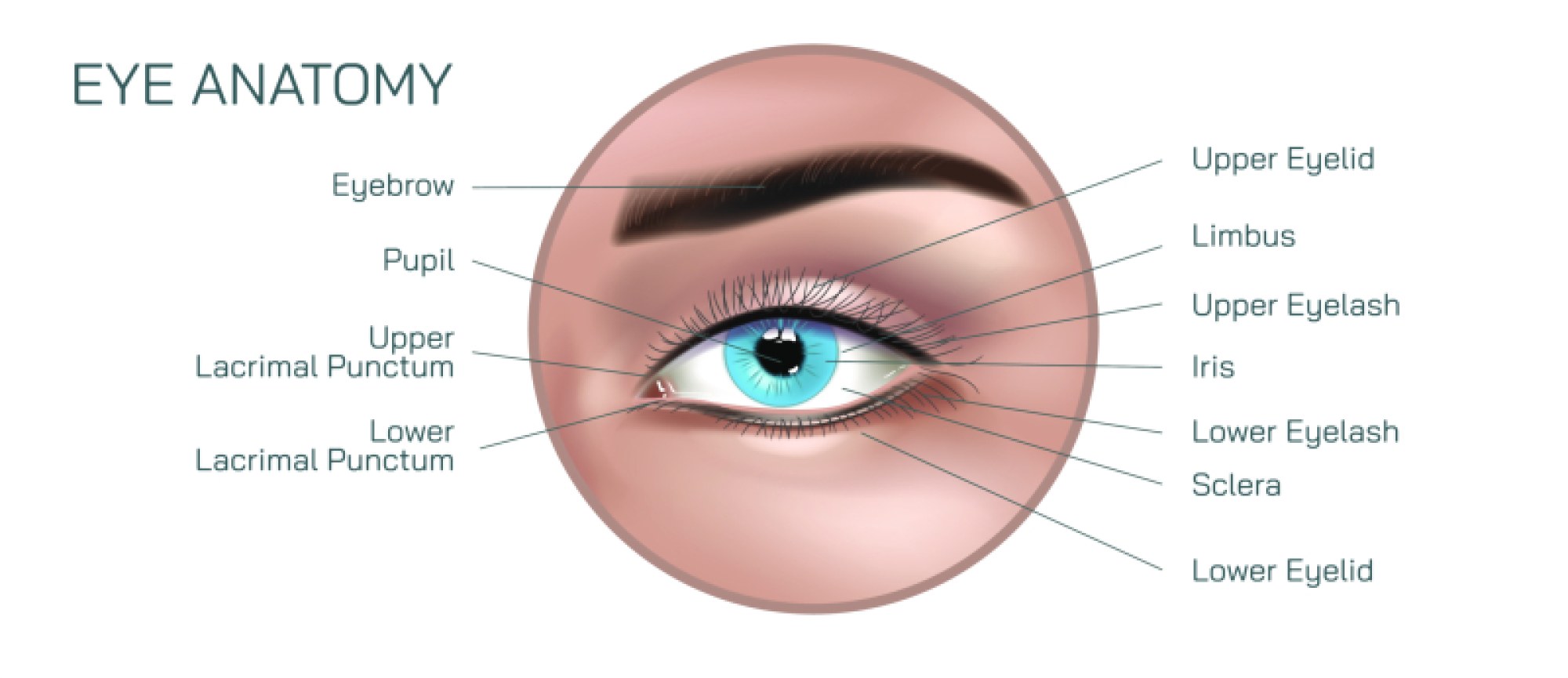
Hypermetropia vs Myopia Illustration: Farsightedness and Nearsightedness Eye Defects Explained
Hypermetropia (farsightedness) and myopia (nearsightedness) are two of the most common refractive vision defects in humans, and an illustration comparing them brings clarity to how the eye focuses light and how subtle differences in eye shape dramatically affect visual accuracy. Although both conditions stem from a mismatch between the optical power of the eye and the length of the eyeball, their effects are opposite: one makes distant objects clear but near objects blurry, while the other makes near objects clear but distant objects blurry. When visualized through diagrams that trace light rays entering the eye and arriving at different focal points, the differences become intuitive and easy to understand.
In a normal (emmetropic) eye, incoming light rays from the environment pass through the cornea and lens and converge precisely on the retina—specifically on the fovea, the region responsible for sharp central vision. Illustrations of normal vision often show light rays traveling through the cornea and lens and forming a crisp focal point right on the retinal surface. The lens adjusts shape through accommodation to fine-tune focus for objects at different distances, allowing the eye to maintain clarity whether the person looks near or far. This balanced function contrasts sharply with what is seen in refractive errors.
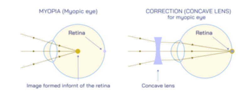
In myopia (nearsightedness), the eye focuses light in front of the retina instead of directly on it. This happens most commonly because the eyeball is too long, or because the cornea has excess curvature that gives the eye too much refractive power. Illustration of myopic vision shows parallel light rays entering the eye and coming to a focal point just before they reach the retina. Because of this, distant objects appear blurry, while near objects are seen clearly, since light from nearer objects diverges and requires less focusing power. A myopic eye illustration often depicts a stretched eyeball with an elongated axial length. Corrective visuals include concave (minus/–) lenses, drawn as diverging lenses placed in front of the eye; these lenses spread out incoming light slightly so that the cornea and lens combine to focus the rays further back—precisely onto the retina.
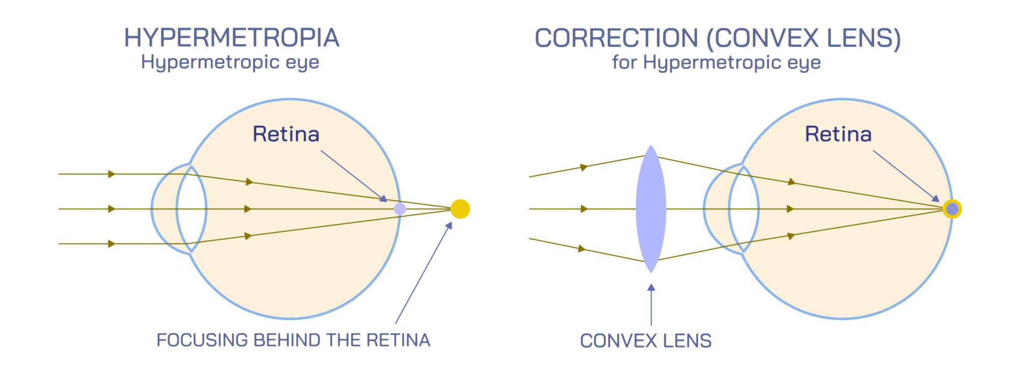
In hypermetropia (hyperopia or farsightedness), the eye focuses light behind the retina. This defect usually occurs because the eyeball is too short, or the cornea has insufficient curvature, giving the eye too little refractive power. The illustrated light-path of a hypermetropic eye shows rays entering and remaining slightly unfocused, converging at a point beyond the retinal surface. As a result, near objects appear blurry, especially when focusing demands exceed the eye’s accommodation abilities. Distant objects are often seen clearly, at least in younger individuals, because the lens can adjust shape to increase focusing power. Illustrations also often include a depiction of the ciliary muscles working harder to thicken the lens for near focus, explaining why people with untreated hypermetropia experience headaches, eye strain, and visual fatigue when reading or doing close work. Corrective visuals show convex (plus/+) lenses, which converge light slightly before it enters the eye so that the retina becomes the true focal plane.
A side-by-side illustration comparing myopic and hypermetropic eyes usually highlights the opposing focal errors visually:
• In myopia, the focal point lies in front of the retina.
• In hypermetropia, the focal point lies behind the retina.
These comparative diagrams underline that the issue is not blurred images themselves but where light converges in relation to the retinal layer.
Some diagrams expand further to show accommodation differences between the two refractive errors. In myopia, accommodation cannot shift the focal point far enough backward to see distant objects clearly. In hypermetropia, accommodation may temporarily compensate for the defect by pushing the focal point forward toward the retina, though this becomes difficult with age. This visual comparison explains why children and young adults with mild hypermetropia can sometimes see clearly without glasses but develop symptoms of eye strain—and why adults often notice worsening near vision as natural accommodation weakens over time.
Illustrations sometimes connect these refractive errors to anatomical risk factors and long-term complications. Myopia, especially high myopia, may be associated with increased risk of retinal detachment, glaucoma, and macular degeneration due to the elongated shape of the eye stretching ocular tissues. Illustrations of hypermetropia may show a shallow anterior chamber and highlight the increased risk of angle-closure glaucoma. These additions help reinforce the fact that refractive defects are not always cosmetic concerns but may relate to deeper ocular health implications.
Enhanced visual learning diagrams may also include laser and lens-based correction techniques. Myopia is often corrected by reshaping the cornea so that its refractive power is reduced, while hypermetropia requires increasing corneal curvature. In severe cases, lens implants or refractive lens exchange may be used. While these surgical concepts extend beyond standard refractive anatomy, showing them visually next to optical correction diagrams reinforces how understanding the focal path guides both glasses prescriptions and medical interventions.
Ultimately, an illustration comparing hypermetropia and myopia does more than depict blurry vision—it provides a visual explanation of how eye shape, corneal curvature, lens power, and accommodative ability determine whether light strikes the retina correctly. It demonstrates that clear vision depends on precise focusing, and when that precision shifts forward or backward relative to the retina, the eye compensates with glasses, contact lenses, or surgical correction. By turning invisible optical principles into clear images, the illustration helps viewers see the eye not just as an organ of sight but as a finely calibrated optical system—one whose accuracy determines whether the world appears crisp from afar, sharp up close, or both.