Human Skin Layers Illustration: Epidermis, Dermis, and Hypodermis Structure Explained
The human skin is the largest organ of the body and an illustration that displays its three major layers—the epidermis, dermis, and hypodermis—reveals how this protective covering functions not only as a barrier but also as an active, living interface between the body and the external world. While skin may appear from the outside as a smooth and uniform surface, a cross-section illustration shows it as a multilayered system filled with cells, fibers, glands, nerves, blood vessels, and adipose tissue, all working together to regulate temperature, provide sensation, support immunity, and maintain hydration. The complexity embedded within these layers highlights why skin is more than an envelope of tissue—it is a dynamic organ essential to survival.
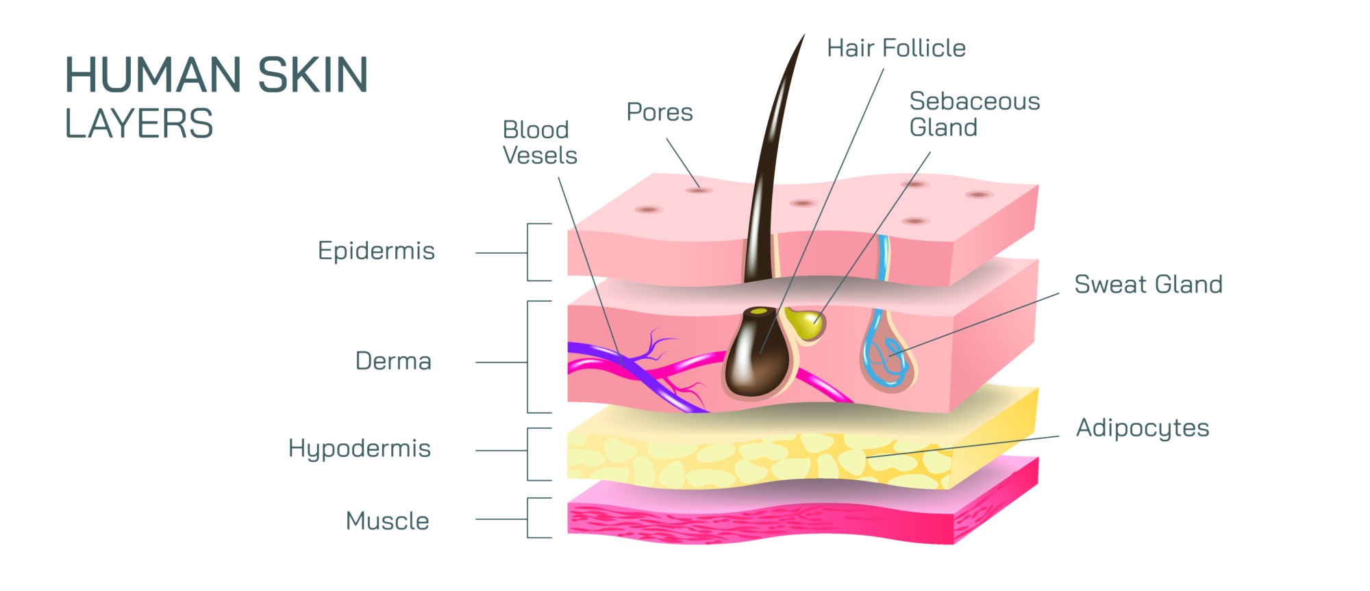
At the very top of the illustration sits the epidermis, the outermost layer of the skin and the body’s first line of defense against the environment. It is a stratified structure composed mainly of keratinocytes, the cells that produce keratin—a tough, water-resistant protein that forms a protective shield. Detailed diagrams show the epidermis organized into distinct sublayers that transition from deep, living cells to flattened dead cells at the surface. The deepest sublayer, the stratum basale, contains actively dividing keratinocytes, supplying new cells that gradually migrate upward. This layer also houses melanocytes, the pigment-producing cells responsible for skin color and protection from ultraviolet radiation. Above this lies the stratum spinosum, where cells interlock through desmosomes, creating structural strength. The next layer, the stratum granulosum, begins the transformation in which cells become flattened, lose their nuclei, and accumulate keratohyalin granules that enable keratin formation. In thicker skin—such as the palms and soles—the stratum lucidum forms an additional translucent band, providing extra resistance to friction. The uppermost stratum corneum is composed of multiple layers of dead, flattened keratinized cells tightly packed like shingles. Illustrations often show these cells sloughing off naturally, symbolizing continuous renewal.
While the epidermis is essential for barrier protection, it does not contain blood vessels and instead relies on the underlying dermis for nourishment. The dermis is shown in illustrations as a thicker, fibrous middle layer rich in collagen and elastin fibers, which make skin both strong and flexible. It is subdivided into two interconnected regions: the papillary dermis and the reticular dermis. The papillary dermis lies immediately beneath the epidermis and features loose connective tissue, capillaries, and sensory receptors. Fingerlike projections known as dermal papillae interlock with the epidermis to secure the layers together and increase surface area for nutrient exchange. The reticular dermis, a deeper and denser region of connective tissue, houses the major structural components of the skin: thick collagen bundles that resist tearing, elastin fibers that enable stretch and recoil, and fibroblasts that produce these proteins. In visual diagrams, the reticular dermis appears as a thick woven matrix that reinforces how the skin withstands mechanical pressure from movement, lifting, gripping, and impact.
A rich network of blood vessels and nerves runs throughout the dermis, and illustrations commonly emphasize their placement. Capillaries near the surface play a crucial role in thermoregulation by dilating to release heat or constricting to conserve it. Sensory nerve endings and specialized receptors—such as Meissner’s corpuscles for light touch, Pacinian corpuscles for deep pressure and vibration, and free nerve endings for pain—are labeled to show how the skin interprets temperature, texture, and injury. Illustrations also highlight hair follicles, which originate deep within the dermis and extend up through the epidermis. Each follicle is associated with a sebaceous gland that produces sebum to moisturize skin and hair, along with an arrector pili muscle capable of contracting to produce goosebumps. Sweat glands—both eccrine glands that regulate temperature and apocrine glands associated with scent in specific body regions—are drawn as coiled structures seated deep in the dermis with ducts running toward the skin surface. Such glandular mapping reinforces that temperature control, lubrication, and odor production are the result of dermal organ systems, not the surface alone.
Beneath the dermis lies the hypodermis, also known as the subcutaneous layer or superficial fascia, and diagrams portray it as a thick layer composed mainly of adipose (fat) tissue, connective tissue, and larger blood vessels. This layer insulates the body from temperature extremes, cushions internal organs from impact, and serves as a major energy reserve. The adipocytes—round, lipid-filled cells—are often illustrated densely packed in clusters that form the soft contour of the body. The hypodermis plays a crucial role in anchoring the skin to underlying muscles and bones through fibrous connective tissue compartments. Its thickness varies depending on anatomical location, age, sex, and nutritional status. Areas such as the abdomen, thighs, and buttocks typically contain a well-developed hypodermis, whereas the eyelids and shins contain very little. In medical illustrations, this layer is also shown as the injection target for subcutaneous medications because of its slow absorption properties.
A complete skin-layer illustration also demonstrates how the three layers work together as a unified organ despite their structural differences. When the body experiences heat, moisture, cold, abrasion, or infection, the epidermis, dermis, and hypodermis respond cooperatively. For example, during heat stress, eccrine sweat glands in the dermis increase sweat production while superficial blood vessels dilate to assist cooling. In cold environments, blood vessels constrict and the hypodermal fat preserves body heat. During injury, the epidermis launches rapid cell turnover while the dermis mobilizes fibroblasts and immune cells to repair connective tissue and rebuild structural fibers. Illustrations that trace wound-healing stages show scab formation, inflammation, collagen deposition, and remodeling—highlighting layers involved at each stage.
The skin is also a sensory and immune organ, and some diagrams visualize immune cells such as Langerhans cells located in the epidermis and dermal immune surveillance cells that detect pathogens or allergens. These visual elements demonstrate how the skin participates in early immune responses, not just passive protection. Likewise, hydration and barrier illustrations often display lipid matrix structures between epidermal cells, explaining how moisture retention depends on intact skin barriers—and why dryness, eczema, or cracking occurs when the barrier is damaged.
Additional versions of this illustration show regional differences in skin thickness across the body. Skin over the heels and palms contains a much thicker stratum corneum to withstand friction, while skin over the eyelids is extremely thin to allow sensitive movement. Illustrations comparing these locations highlight that the layered structure of skin is consistent everywhere but varies proportionally based on the mechanical demands of that body region.
From a developmental perspective, diagrams sometimes compare skin across the lifespan, showing that infants have thinner epidermis and dermis with highly permeable skin, while aging skin gradually loses collagen, elastin, hydration, and thickness. These comparisons reveal how wrinkles, sagging, dryness, and slower wound healing result from structural changes deep within the layers rather than only surface-level alterations.
Ultimately, an illustration of the epidermis, dermis, and hypodermis transforms the skin from something we see only externally into a layered biological system with overlapping roles in protection, sensation, temperature control, fluid balance, immune defense, metabolism, and structural support. By showing cells dividing in the epidermis, fibers strengthening the dermis, and fat cushioning the hypodermis, the illustration turns skin into an understandable organ—one whose complexity supports virtually every aspect of human function and daily life.