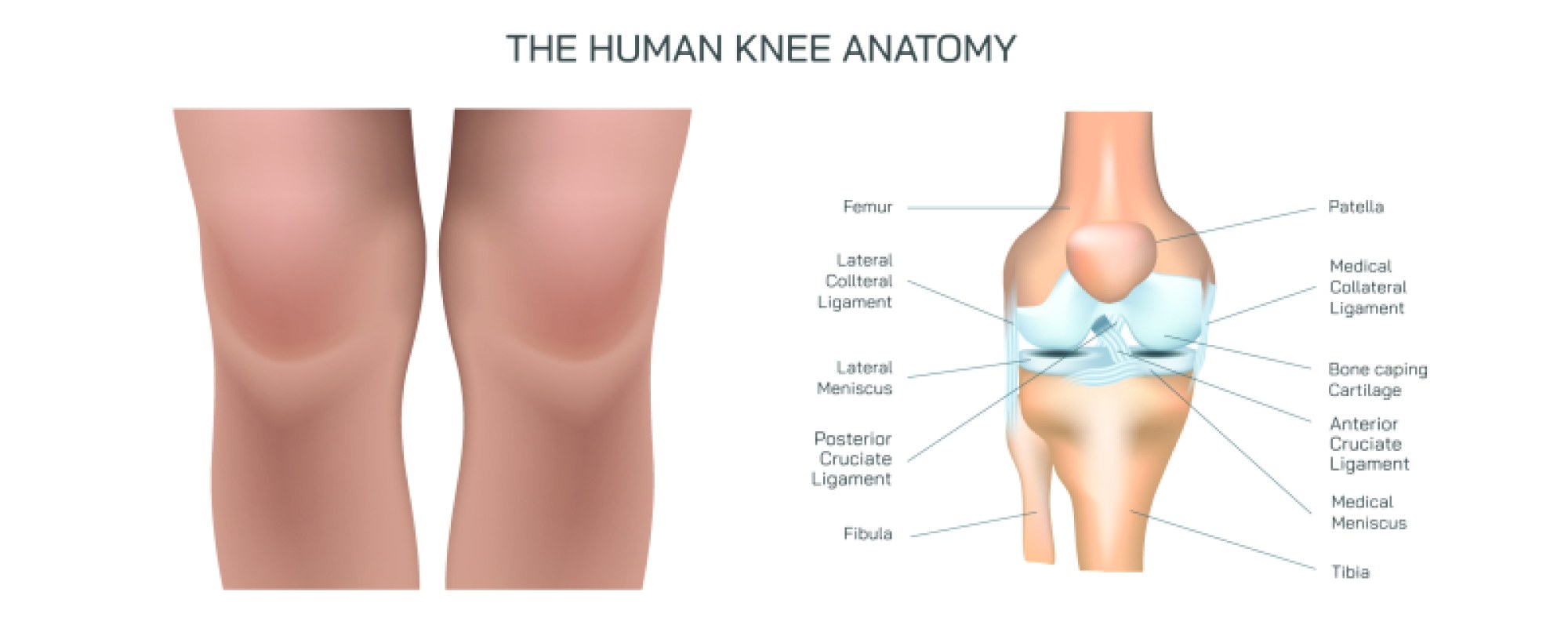
Placentation of Twins in the Human Womb Illustration: Monozygotic and Dizygotic Placental Structures Explained
Twins represent one of the most captivating biological variations in human reproduction, and an illustration showing placentation—how the placenta and fetal membranes form around developing twins—reveals not only the different ways multiple pregnancies begin but also how early embryologic events influence the structure, risks, and clinical outcomes of twin gestation. Although all pregnancies begin with the implantation of a fertilized egg into the uterine lining, twin pregnancies diverge along two fundamental biological pathways. One scenario involves dizygotic (fraternal) twins, in which two separate eggs are fertilized by two separate sperm during the same cycle, producing genetically unique siblings who share the womb. The other involves monozygotic (identical) twins, in which a single fertilized egg splits into two embryos. Whether twins are dizygotic or monozygotic determines more than their genetic similarity—it also strongly influences the type of placentation: the number of placentas, the number of amniotic sacs, and the degree to which blood circulation is shared. A clear illustration makes these relationships immediately visible by depicting the uterine cavity and showing how the number of placentas and membranes varies depending on when twin embryos separate.
Dizygotic twins are straightforward in placental diagrams because they almost always implant independently. Each embryo forms its own placenta and its own chorionic and amniotic sacs, resulting in a dichorionic–diamniotic (Di/Di) pregnancy. In a visual representation, dizygotic twins appear positioned separately in the uterus, each enclosed in its own amniotic sac and surrounded by its own chorion, with the two placentas either separated completely or joined superficially if implantation occurs close together. Even when dizygotic placentas appear fused in drawings or ultrasounds, they remain physiologically separate. The thick dividing membrane between the twins represented in the illustration consists of four layers: two amnions and two chorions, signifying complete separation of support systems. Clinically, this configuration carries the lowest risk among multiple pregnancies because each fetus controls its own environment and blood flow.
Monozygotic twins require a more complex illustration because their placentation depends on the timing of embryonic splitting. When a single fertilized egg divides within the first 2–3 days after fertilization, before the chorion has begun to form, the outcome resembles dizygotic twins structurally: each twin develops with its own placenta, chorion, and amnion—a dichorionic–diamniotic (Di/Di) monozygotic pregnancy. This arrangement appears identical to dizygotic twins on a diagram, making placentation alone insufficient for distinguishing genetic identity; genetic testing, sex identification, or later imaging features must be used. When splitting occurs slightly later, between days 4 and 8, after the chorion begins developing but before the amnion forms, the placentation becomes monochorionic–diamniotic (Mo/Di). In this configuration, one placenta supports both twins, there is one shared chorion, but two separate amniotic sacs, each enclosing a fetus individually. Ultrasound and anatomical illustrations display a single placenta connected to both umbilical cords and a thinner membrane dividing the twins, made up of only two amniotic layers without chorionic separation. Mo/Di twins are the most common form of monozygotic twinning and carry moderate clinical risk due to their shared placental circulation.
If splitting takes place later still—between days 8 and 13, after both the chorion and the amnion have formed—the result is monochorionic–monoamniotic (Mo/Mo) twins. In this highest-risk arrangement, the illustration shows one placenta, one chorion, and a single shared amniotic sac, with no membrane dividing the twins. The umbilical cords float freely in the same sac, which is why the risk of cord entanglement, reduced blood flow, and compression increases significantly. Mo/Mo twins are rare but medically monitored very closely, and visual diagrams emphasize features such as the absence of a dividing membrane and the proximity of the fetuses within the same fluid space. Finally, in the rarest and latest separation—occurring after day 13—splitting is incomplete, resulting in conjoined twins, who appear in illustrations physically fused at one or more anatomical regions while still sharing a placenta and amniotic environment. This depiction reinforces how placentation reveals the developmental timing of embryo separation in monozygotic twins.
One of the most crucial structures highlighted in placentation illustrations of monochorionic twins is the shared vasculature within the placenta. Multiple vascular connections—artery-to-artery, artery-to-vein, or vein-to-vein—form natural anastomoses that allow blood to flow between the twins. Although such networks can equalize circulation in some cases, they also underlie the development of Twin-to-Twin Transfusion Syndrome (TTTS), in which blood flow becomes imbalanced, causing one twin (the donor) to experience reduced blood volume and amniotic fluid while the other (the recipient) receives excess blood and amniotic fluid. Clinical diagrams show this imbalance with arrows illustrating the direction of transfusion between shared arteries and veins, often accompanied by dramatic differences in fluid volume and fetal size. Such diagrams highlight the need for monitoring monochorionic twins closely during pregnancy to detect early signs of circulatory mismatch.
Anatomical illustrations of the fetal membranes deepen understanding of the distinctions between placentation types. The amnion appears as the inner, delicate membrane tightly surrounding the fetus and amniotic fluid, while the chorion forms the outer membrane, rich in vasculature and interfacing directly with the placenta. Di/Di pregnancies show these membranes doubled between fetuses; Mo/Di pregnancies show only the dual amnion dividing them; Mo/Mo pregnancies show neither because both twins occupy a shared amniotic cavity. A clear diagram often labels membrane layers explicitly so viewers can understand how ultrasound imaging distinguishes twin types: Mo/Di twins exhibit the “T-sign” at the membrane–placenta junction, while Di/Di twins show the thicker “lambda sign” caused by chorionic projection between placentas.
The placenta itself—illustrated with its disc-like appearance, branching villi, and deep embedding into the uterine lining—plays an essential role for every fetus but becomes even more influential in twins. It performs nutrient transfer, waste removal, oxygen exchange, immune protection, and hormonal support. In Di/Di pregnancies, each placenta regulates these functions independently, while in monochorionic pregnancies, one organ must balance the needs of two. In diagrams showing umbilical cord insertion sites, unequal placement on the shared placenta may predispose one twin to better perfusion than the other, contributing to selective intrauterine growth restriction (sIUGR)—a condition also commonly depicted in Mo/Di placentation images.
Illustrations of twin placentation also reflect practical obstetric implications. Placentation type influences ultrasound scheduling, delivery planning, risk management, and neonatal care. Di/Di twins are generally monitored less frequently; Mo/Di twins receive more imaging due to risks associated with shared circulation; Mo/Mo twins require the most frequent monitoring, often hospitalization in late pregnancy due to cord-entanglement dangers. Visual timelines and monitoring charts sometimes accompany placentation diagrams to show progressive changes from implantation to full gestation.
Ultimately, illustrations of placentation in twins offer more than a visual guide to the physical structures of the womb—they tell the chronological story of early embryonic development, reveal why twin pregnancies differ so widely in their medical characteristics, and highlight how the placenta acts not merely as a passive attachment but as an active partner sustaining fetal life. By showing how the number of placentas, chorions, and amniotic sacs depends on twinning type and timing, the diagram transforms abstract embryology into a clear understanding of biological structure, clinical risk, and developmental significance. Through these images, one sees how two lives can form side by side—sometimes completely separate, sometimes intricately interconnected—all beginning from the earliest organizational decisions of cells within the womb.