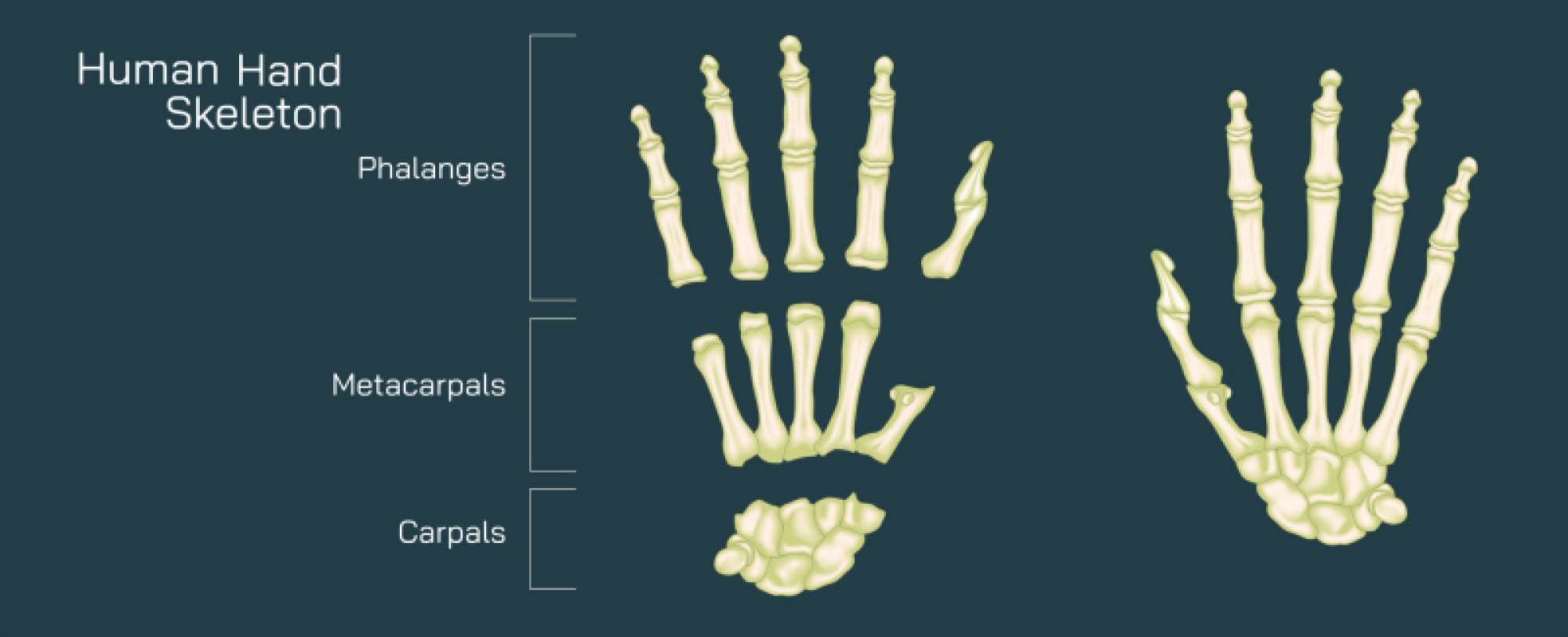
Hand Skeleton Anatomy Vector Illustration Showing Bones of Palm, Fingers, Wrist, and Joint Structure
The human hand is one of the most intricate and versatile structures of the human body, combining precision, strength, and mobility to perform a vast range of functions. Its skeletal anatomy underpins every movement, providing the framework for gripping, manipulating objects, and executing fine motor tasks. A vector illustration of the hand skeleton typically highlights the bones of the wrist (carpals), palm (metacarpals), fingers (phalanges), and the complex joint structures, offering a comprehensive view of both the anatomical layout and functional interconnections. Such diagrams serve as essential tools in medical education, physiotherapy, surgery, and biomechanics, allowing learners to understand the relationship between bones, joints, and movement mechanics in the hand.
The wrist or carpal bones are depicted as a cluster of eight small, irregularly shaped bones arranged in two rows, providing a stable yet flexible base for hand movement. The proximal row, nearest the forearm, includes the scaphoid, lunate, triquetrum, and pisiform, while the distal row, closer to the palm, comprises the trapezium, trapezoid, capitate, and hamate. Vector illustrations often label each carpal bone individually and may highlight their articulations with the radius and ulna of the forearm, demonstrating how the wrist allows both flexion-extension and radial-ulnar deviation. The arrangement of the carpals in two rows also facilitates the transmission of forces from the hand to the forearm, ensuring both stability and adaptability during gripping or weight-bearing activities.
Extending from the wrist are the metacarpal bones, five long bones forming the framework of the palm. Each metacarpal connects proximally to the distal row of carpal bones and distally to the proximal phalanges of the fingers. In vector illustrations, metacarpals are often numbered from one to five, beginning with the thumb side (lateral aspect) to the little finger (medial aspect). The diagram may include annotations showing the base, shaft, and head of each metacarpal, emphasizing their structural differences and functional roles. The first metacarpal, supporting the thumb, is shorter and more mobile, allowing opposition—a key feature enabling grasping and fine motor tasks.
The phalanges of the fingers are typically depicted in three segments for each finger—proximal, middle, and distal phalanges—except the thumb, which has only two. Vector illustrations often show the alignment of these bones and the formation of the finger joints, including metacarpophalangeal (MCP), proximal interphalangeal (PIP), and distal interphalangeal (DIP) joints. Arrows or labels may indicate joint movements such as flexion, extension, abduction, and adduction. This visual representation highlights the functional range of motion and the biomechanical interactions that allow dexterity, from grasping large objects to manipulating tiny tools. Color coding or shading can differentiate between bone types or joint areas, enhancing comprehension of complex articulations.
The joint structure of the hand is crucial for coordinated movement. The vector illustration may depict the carpometacarpal (CMC) joints connecting the metacarpals to the carpal bones, the MCP joints linking the metacarpals to the proximal phalanges, and the interphalangeal joints facilitating bending and straightening of the fingers. The thumb’s saddle-shaped CMC joint is often highlighted to illustrate its unique contribution to opposition and precision grip. Ligament attachments, while sometimes simplified, can be represented to show how stability is maintained across joints, preventing dislocations while allowing mobility.
Vector illustrations also often incorporate skeletal alignment from multiple views—palmar, dorsal, and lateral—to provide a complete understanding of spatial orientation. Labels indicate not only individual bones but also articulating surfaces and joint spaces, helping learners visualize how bones interact during movement. Magnified sections may highlight the articulations of smaller bones, such as the scaphoid and trapezium, which are particularly prone to injury.
Additional elements in the vector diagram may include clinical references, showing areas susceptible to fractures, such as the scaphoid in the wrist or the distal phalanges in the fingers. Highlighting these regions can provide practical context for medical and physiotherapy education, emphasizing the importance of anatomy in diagnosing and treating hand injuries. Some illustrations may also indicate muscle attachment points on bones, helping connect skeletal structure with functional movements powered by extrinsic and intrinsic hand muscles.
Color coding is frequently used in vector illustrations to distinguish between wrist, palm, and finger bones, enhancing clarity in complex diagrams. Arrows may show the flow of forces or the direction of movements at different joints, integrating anatomical structure with biomechanical function. Labels and legends ensure that each bone and joint is identifiable, making the illustration suitable for learning, reference, and professional practice.
By combining carpal, metacarpal, and phalangeal bones with joint articulations and spatial orientation, a hand skeleton vector illustration provides a thorough understanding of the structural foundation of hand movement. It highlights the interdependence of bones and joints, the unique anatomy of the thumb, and the intricate arrangement that allows the hand to perform both powerful and delicate tasks. Such diagrams are indispensable in medical education, rehabilitation, ergonomic design, and any discipline that requires detailed knowledge of hand anatomy.
Ultimately, a vector illustration of the hand skeleton demonstrates the complexity and elegance of human anatomy. It conveys how the bones of the wrist, palm, and fingers, in conjunction with joint structures, provide both stability and dexterity. By integrating detailed labeling, multiple perspectives, and functional insights, the diagram transforms a complex anatomical system into an educational and visually intuitive framework, enabling comprehensive understanding of the human hand’s skeletal architecture.