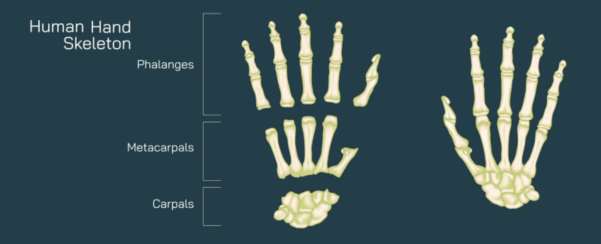
Hand Skeleton Anatomy Vector Illustration Showing Bones, Joints, and Structure of Human Hand for Medical and Educational Study
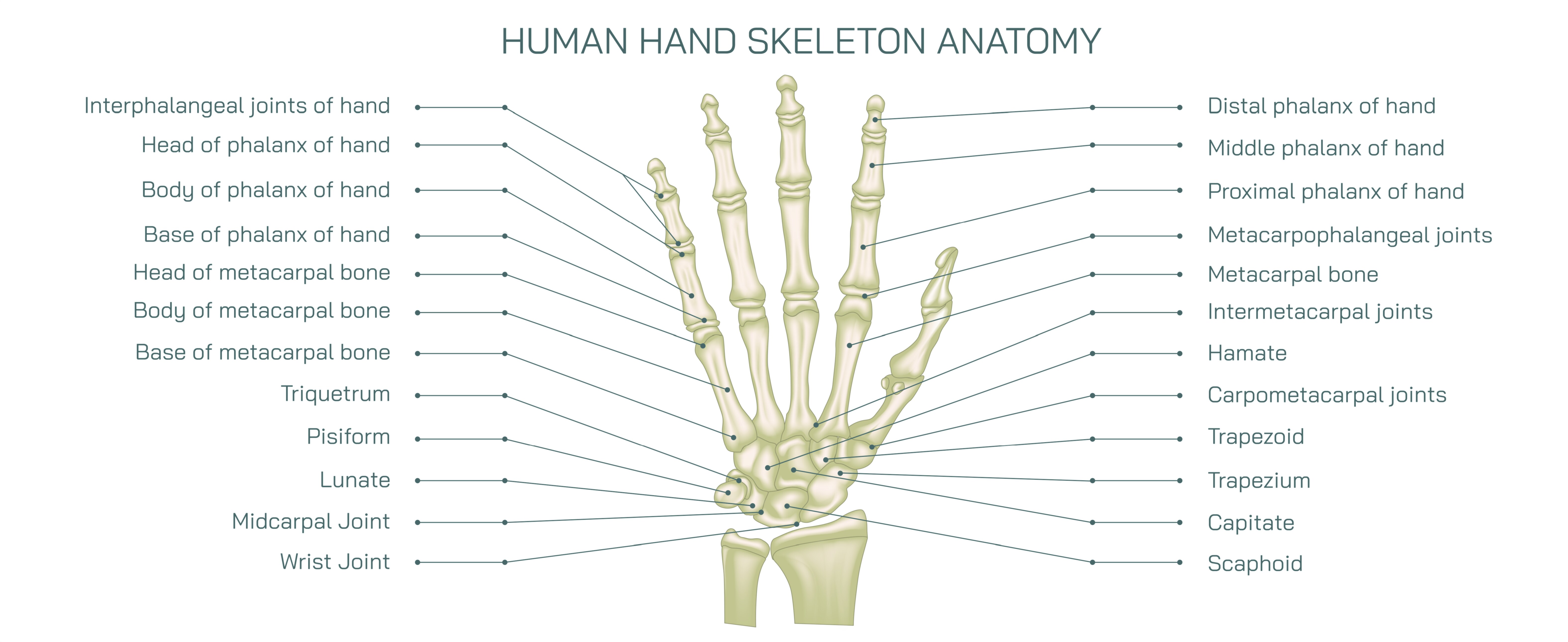
The human hand is a highly intricate structure, combining bones, joints, muscles, ligaments, and tendons to provide remarkable dexterity, strength, and precision. Understanding the skeletal anatomy of the hand is essential for medical education, orthopedic practice, physiotherapy, and anatomy studies. A vector illustration of hand skeleton anatomy typically integrates all bones, joints, and articulations with clear labeling, providing a detailed and educational depiction of the structural framework that supports hand function. By combining dorsal and palmar perspectives, color-coded bones, and directional annotations, such illustrations make complex anatomical relationships visually accessible.
At the center of the illustration is the overall hand skeleton, often shown in a slightly angled perspective to provide depth and clarity. The hand is divided into three main regions: the carpals (wrist bones), metacarpals (palm bones), and phalanges (finger bones). Color coding is typically used to distinguish these regions, with carpals in one hue, metacarpals in another, and phalanges in a third. Arrows and labels indicate bone names, articulations, and orientation, providing immediate visual cues for anatomical identification.
The carpal bones are depicted as two rows of short, irregular bones forming the wrist. The proximal row includes the scaphoid, lunate, triquetrum, and pisiform, while the distal row includes the trapezium, trapezoid, capitate, and hamate. Vector diagrams often include magnified insets showing individual carpal bone shapes and labeling articulations with the radius and ulna proximally, and the metacarpals distally. Arrows may indicate rotational movement and flexion-extension axes of the wrist, highlighting functional articulation.
The metacarpals, forming the palm, are depicted as five long bones connecting the carpals to the phalanges. Each metacarpal is labeled numerically from I (thumb) to V (little finger), with arrows indicating their alignment relative to the carpal bones and phalanges. Magnified insets may illustrate the metacarpophalangeal (MCP) joints, highlighting their role in finger flexion, extension, abduction, and adduction. Color coding or shading differentiates metacarpal heads, shafts, and bases for clarity.
The phalanges, forming the fingers, are depicted with three bones for each finger (proximal, middle, distal) and two for the thumb (proximal and distal). Labels identify each phalanx, while arrows indicate joint types: proximal interphalangeal (PIP), distal interphalangeal (DIP), and thumb interphalangeal joints. Additional arrows may indicate flexion-extension movement axes, demonstrating how the skeletal framework supports finger dexterity. Color differentiation or gradients can highlight the phalangeal regions, while overlays indicate joint surfaces and articulations.
Vector illustrations often include joint labeling, showing carpometacarpal (CMC), metacarpophalangeal (MCP), and interphalangeal (IP) joints, along with key ligaments such as collateral ligaments. Arrows may indicate joint movement or axes of rotation, linking skeletal anatomy with functional motion. Insets may magnify thumb CMC joint anatomy, emphasizing its unique saddle configuration and role in opposability.
Additional features often include palmar and dorsal perspectives. The palmar view may highlight the alignment of metacarpals and phalanges relative to hand arches, while the dorsal view emphasizes carpal arrangement and extensor tendon pathways. Color coding and labeled arrows help distinguish dorsal versus palmar bones, providing comprehensive spatial understanding.
Vector diagrams may also include clinical or educational annotations, highlighting landmarks for bone palpation, fracture sites (e.g., scaphoid fractures), and common joint disorders such as osteoarthritis in MCP or PIP joints. Arrows may indicate typical force transmission pathways from the wrist through the hand during gripping or pinching activities, connecting skeletal anatomy to functional outcomes.
By combining carpals, metacarpals, phalanges, joint articulations, and directional movement axes, a hand skeleton vector illustration provides a detailed, comprehensive view of hand anatomy. Color-coded regions, labeled bones, magnified joint insets, and arrows indicating motion allow learners to understand structural relationships and functional implications simultaneously. Comparative panels may show left versus right hands or palmar versus dorsal perspectives, enhancing educational clarity.
Ultimately, a vector illustration of the human hand skeleton demonstrates the integration of bones and joints that support complex hand movements, linking anatomical structure to functional biomechanics. Through labeled bones, articulated joints, color-coded regions, and directional arrows, the diagram transforms abstract skeletal information into a visually engaging and educational tool, suitable for medical study, orthopedic education, physiotherapy training, and anatomy learning.