Position of Baby Fetus in Uterus Vector Illustration Showing Fetal Alignment, Cephalic and Breech Positions, and Pregnancy Stages
The position of a fetus in the uterus is a critical aspect of maternal and fetal health, as it directly influences labor, delivery outcomes, and obstetric decision-making. Understanding fetal alignment, including cephalic (head-down) and breech (feet- or buttocks-first) positions, and how these positions change during pregnancy, is essential for medical students, obstetricians, midwives, and expectant parents. A vector illustration depicting fetal positions typically combines uterine anatomy, fetal orientation, placental location, and stages of fetal growth, providing a visually intuitive and educational tool. Through labeled structures, directional arrows, and comparative panels, such diagrams clarify complex spatial relationships within the pregnant uterus.
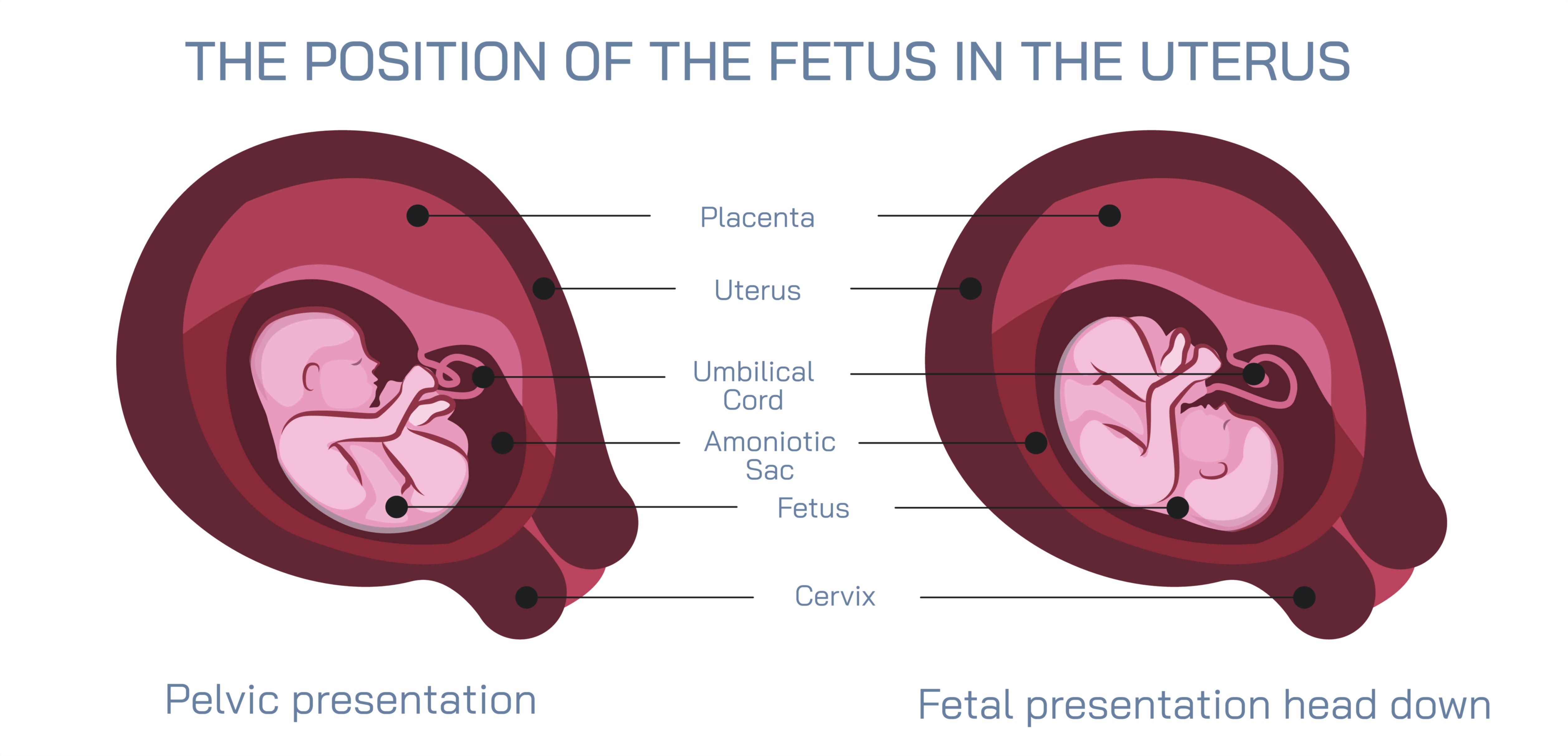
At the center of the illustration is a cross-sectional view of the uterus, showing the myometrium, amniotic sac, placenta, and cervix. The uterus is often depicted in a slightly angled frontal or lateral perspective to enhance visibility of fetal positioning. Color coding is used to differentiate uterine tissue, amniotic fluid, and placenta, while arrows indicate the directional orientation of the fetus relative to the maternal pelvis and birth canal. Labels identify the cervix, uterine fundus, and pelvic bones, providing anatomical context for understanding fetal alignment in late pregnancy.
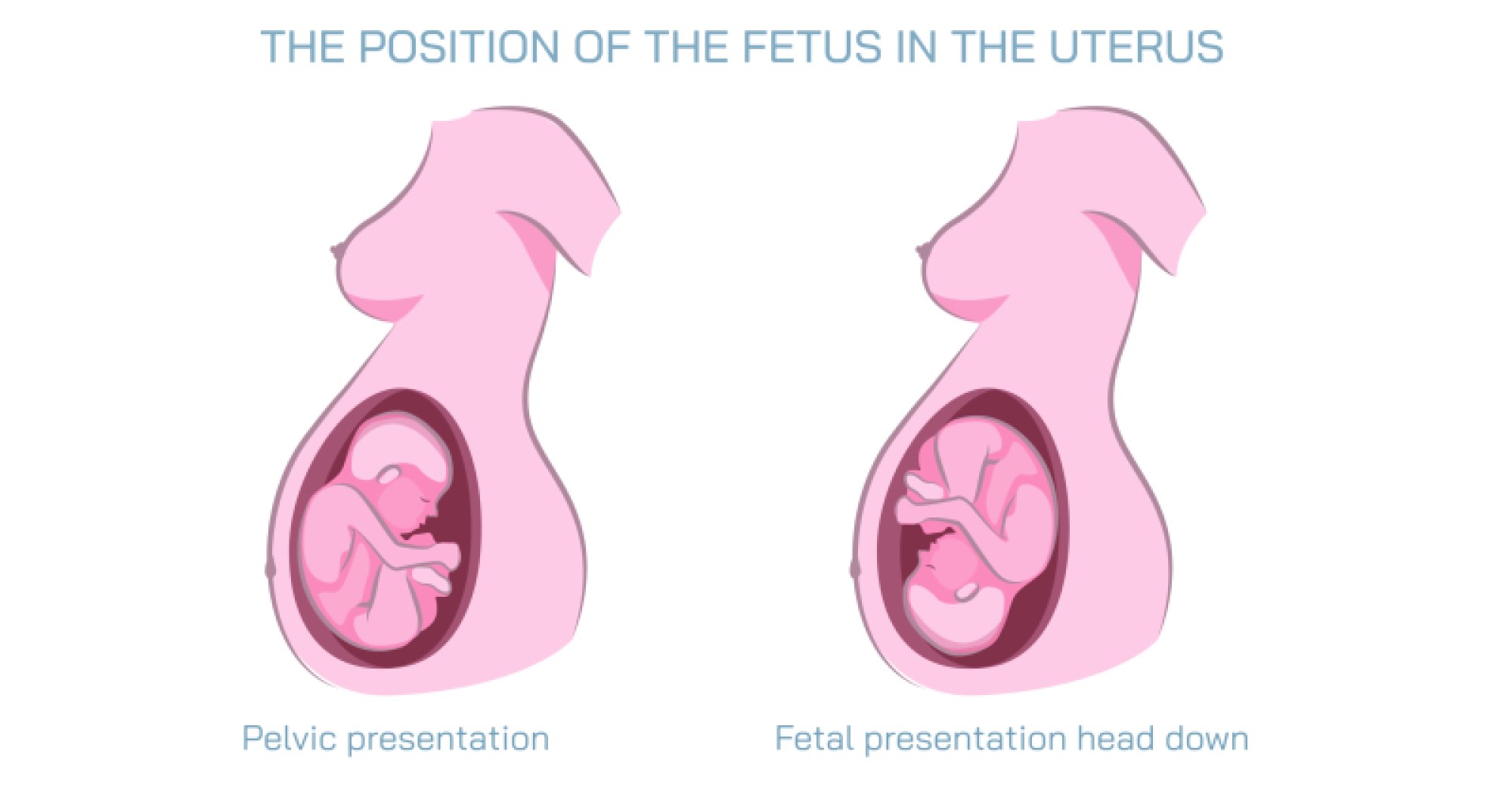
The fetus is depicted inside the uterus, showing accurate anatomical proportions relative to gestational age. For cephalic positions, the fetus is illustrated with its head oriented downward toward the cervix, considered the optimal alignment for vaginal delivery. Subtypes, such as occiput anterior (OA), occiput posterior (OP), and transverse lie, are often indicated, with arrows demonstrating slight rotations or tilts of the fetal head that can impact labor. Labels highlight the fetal head, spine, limbs, and extremities, showing their spatial relationship within the uterine cavity.
Breech positions are depicted with the fetus oriented buttocks or feet downward toward the cervix. Different types of breech are illustrated in separate panels or insets, including frank breech (buttocks-first with extended legs), complete breech (folded legs), and footling breech (one or both feet first). Arrows indicate the direction of fetal orientation, and labels emphasize potential delivery challenges associated with breech presentations, highlighting why early detection is important for obstetric planning. Color coding differentiates breech from cephalic positions, allowing quick visual comparison.
Vector illustrations often include gestational stages, showing how fetal positions evolve from early to late pregnancy. For example, second-trimester diagrams may depict the fetus in more transverse or floating positions due to ample uterine space, while third-trimester illustrations show the fetus increasingly descending into the pelvic cavity with the head engaging the birth canal. Arrows may indicate changes in fetal orientation over time, visually representing dynamic alignment and fetal movement as pregnancy progresses.
Additional features include placental and umbilical cord placement, with labels illustrating normal implantation sites and cord positioning relative to fetal orientation. Magnified insets may depict fetal limb positions, spinal curvature, and head flexion, showing how anatomical alignment affects delivery mechanics. Color-coded shading highlights the fetal body versus surrounding amniotic fluid, enhancing clarity for learners.
Vector diagrams may also include maternal pelvic structures, showing how fetal position interacts with the pelvic inlet and outlet. Arrows may illustrate the pathway the fetus follows during vaginal delivery in both cephalic and breech scenarios. Comparative panels may show optimal versus malpositioned fetuses, reinforcing the importance of prenatal assessment and interventions such as external cephalic version (ECV) or cesarean section when necessary.
By combining uterine anatomy, fetal positions, cephalic and breech variations, gestational stage representation, and maternal pelvic context, a vector illustration provides a comprehensive understanding of fetal positioning. Directional arrows, magnified insets, and color-coded highlights allow learners to distinguish between optimal and non-optimal positions, linking structural knowledge with clinical implications for labor and delivery.
Ultimately, a vector illustration of fetal position in the uterus demonstrates the interplay between fetal alignment, gestational development, and maternal anatomy, visually connecting prenatal growth with obstetric planning and delivery outcomes. Through labeled fetal and uterine structures, directional markers, gestational stage panels, and comparative positioning, the diagram transforms complex spatial and developmental concepts into an intuitive, educational, and visually engaging tool for medical study, patient education, and prenatal care.