Internal and External Eye Anatomy Vector Illustration Showing Human Eye Structure with Labeled Parts for Vision and Medical Study
The human eye is a highly complex sensory organ responsible for capturing light and transmitting visual information to the brain. Understanding its anatomy is essential for medical education, optometry, ophthalmology, and biological studies. A vector illustration of internal and external eye anatomy typically integrates both surface structures and internal components, clearly labeled to depict their relationship to vision and ocular function. By combining external features, internal layers, and directional pathways for light and fluid, such illustrations provide a comprehensive visual framework for learning human eye anatomy.
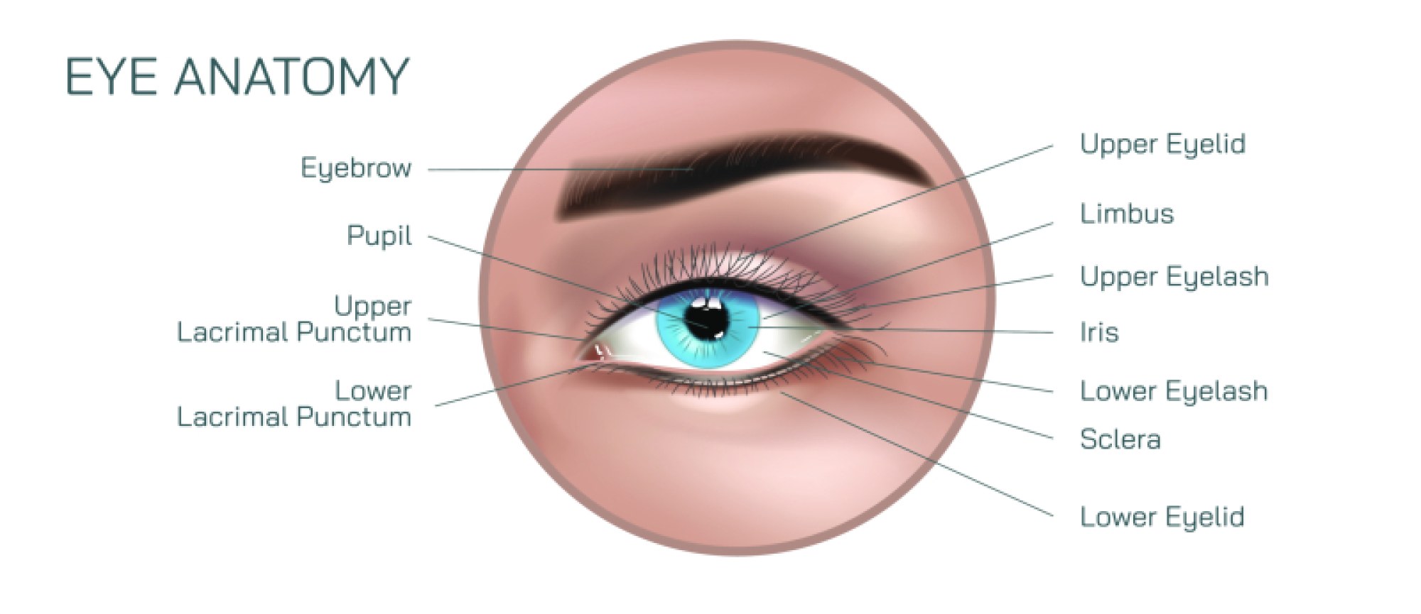
At the center of the illustration is the external anatomy of the eye, often depicted in lateral or frontal view. Key components include the cornea, the transparent dome-shaped structure that refracts incoming light; the sclera, the white, fibrous outer layer that provides structural support; and the limbus, the junction between cornea and sclera. The conjunctiva, a thin membrane covering the sclera, is also labeled to demonstrate its role in protecting the eye from dust and microbes. Additional external structures include the eyelids, eyelashes, and lacrimal apparatus, which maintain moisture and provide protective reflexes. Arrows or magnified insets may illustrate tear production, drainage via the lacrimal ducts, and blink reflex function, highlighting protective and lubrication mechanisms.
The internal anatomy is revealed through a cross-sectional view of the eye. The anterior chamber, filled with aqueous humor, lies between the cornea and the iris, which controls the amount of light entering through the pupil. The lens, a biconvex, flexible structure, focuses light onto the retina by adjusting curvature through ciliary muscle action, a process known as accommodation. Vector diagrams often include arrows showing the light path through the cornea and lens to the retina, visually connecting anatomical features with the functional goal of forming clear images.
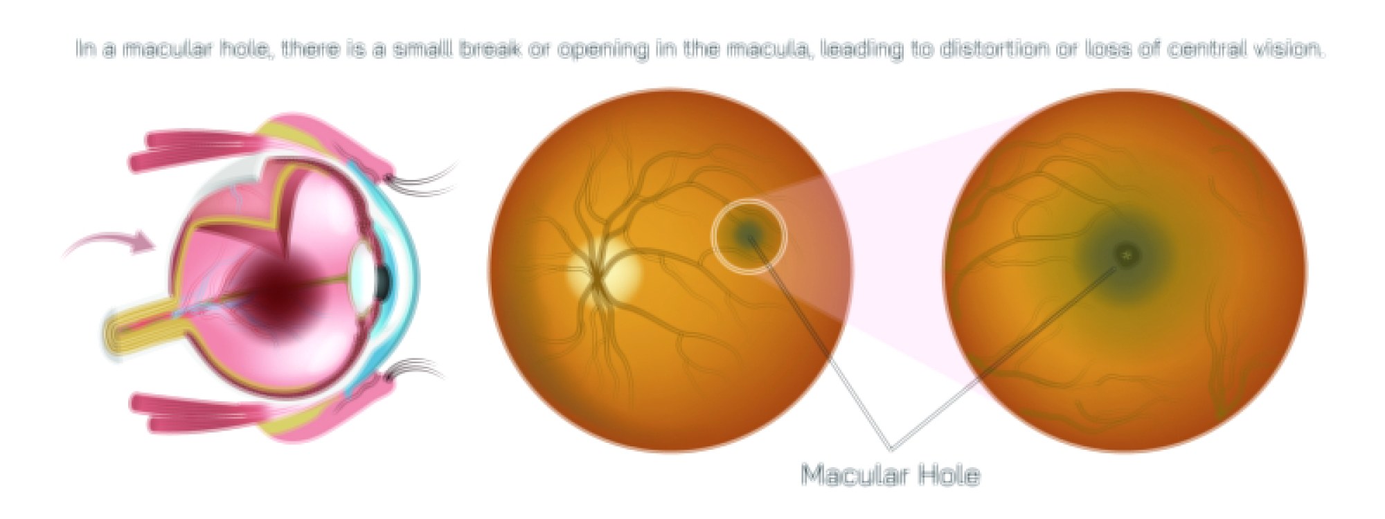
The retina is typically illustrated in detail, highlighting its layered structure. Labeled components include photoreceptor cells—rods for low-light vision and cones for color perception—as well as bipolar cells, ganglion cells, and the optic nerve. Arrows can indicate the flow of visual information from photoreceptors through retinal neurons to the optic nerve, which transmits signals to the brain. Magnified insets may show the macula and fovea, the regions of highest visual acuity, with labels emphasizing their role in detailed central vision.
Vector illustrations also depict the vitreous chamber, filled with the vitreous humor, which maintains eye shape and optical transparency. Labeled anatomical relationships highlight how the vitreous body supports the retina and contributes to intraocular pressure maintenance. The choroid, a vascular layer between the sclera and retina, is often labeled to illustrate its function in supplying oxygen and nutrients to the outer retina. Shading and color coding distinguish fluid-filled chambers, muscular structures, and vascular components, enhancing clarity.
Additional labeled components often include the ciliary body, responsible for lens accommodation and aqueous humor production, and the trabecular meshwork, which facilitates aqueous humor drainage into the canal of Schlemm. The iris pigment and sphincter muscles are shown to demonstrate pupil constriction and dilation, correlating structure with light regulation and visual adaptation. Arrows may indicate fluid movement, muscle contraction, and pupillary responses to changing light conditions.
Vector illustrations may also include anatomical relationships between anterior and posterior segments. For instance, the optic disc, where the optic nerve exits the eye, is depicted along with the blind spot, while labels indicate its lack of photoreceptors. Comparative panels may show a normal cross-section versus an eye with structural anomalies, enhancing educational value for medical study.
Color coding, directional arrows, and magnified insets are commonly used to enhance understanding of complex relationships. External features are typically highlighted in neutral or soft colors, while internal structures are depicted with distinct hues to separate cornea, lens, retina, and vascular layers. Arrows along the light path and fluid movement help learners correlate anatomical structures with physiological functions, reinforcing functional anatomy alongside structural identification.
By combining external structures, anterior and posterior chambers, lens, retina, optic nerve, and accessory components, a vector illustration of the human eye provides a comprehensive overview of both visible and internal anatomy. Labels, magnifications, and directional arrows clarify the roles of each component in vision, fluid regulation, and ocular health. Such diagrams are invaluable for medical students, optometry trainees, ophthalmologists, and educators, providing a visual reference for understanding normal eye structure, optical pathways, and functional relationships.
Ultimately, a vector illustration of internal and external eye anatomy demonstrates the integration of structural and functional features necessary for vision, combining corneal refraction, lens focusing, retinal photoreception, and neural transmission into a cohesive visual framework. Through clearly labeled parts, color-coded chambers, light path arrows, and magnified insets, the diagram transforms complex ocular anatomy into an educational, visually engaging, and intuitive tool for medical study and vision science education.