Rheumatoid Arthritis Joint Inflammation and Autoimmune Disease Illustration
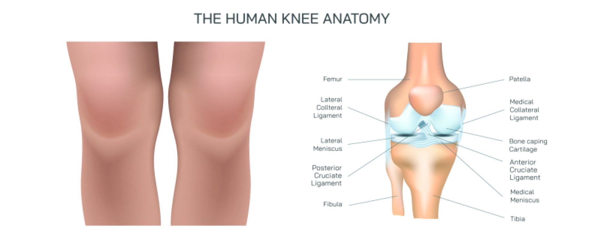
Rheumatoid arthritis is one of the most extensively studied autoimmune diseases because of the profound impact it has on the joints, connective tissues, and daily function of individuals who develop it. A realistic illustration of rheumatoid arthritis centers on the transformation that occurs within a joint—particularly one that appears healthy in its early stages yet slowly becomes a site of persistent inflammation, structural damage, and immune-driven tissue destruction over time. In a normal joint, smooth cartilage cushions the ends of bones and allows friction-free movement while the synovial membrane produces lubricating fluid that maintains the integrity of the joint space. The capsule that surrounds the joint protects these structures from mechanical stress and external damage. In rheumatoid arthritis, however, this entire landscape changes because the immune system mistakenly identifies components of the synovial membrane as foreign and mounts an inflammatory attack against the very tissues it is meant to protect. The result is not a short-lived flare but a prolonged autoimmune response that alters anatomy, function, and mobility while causing significant pain and swelling.
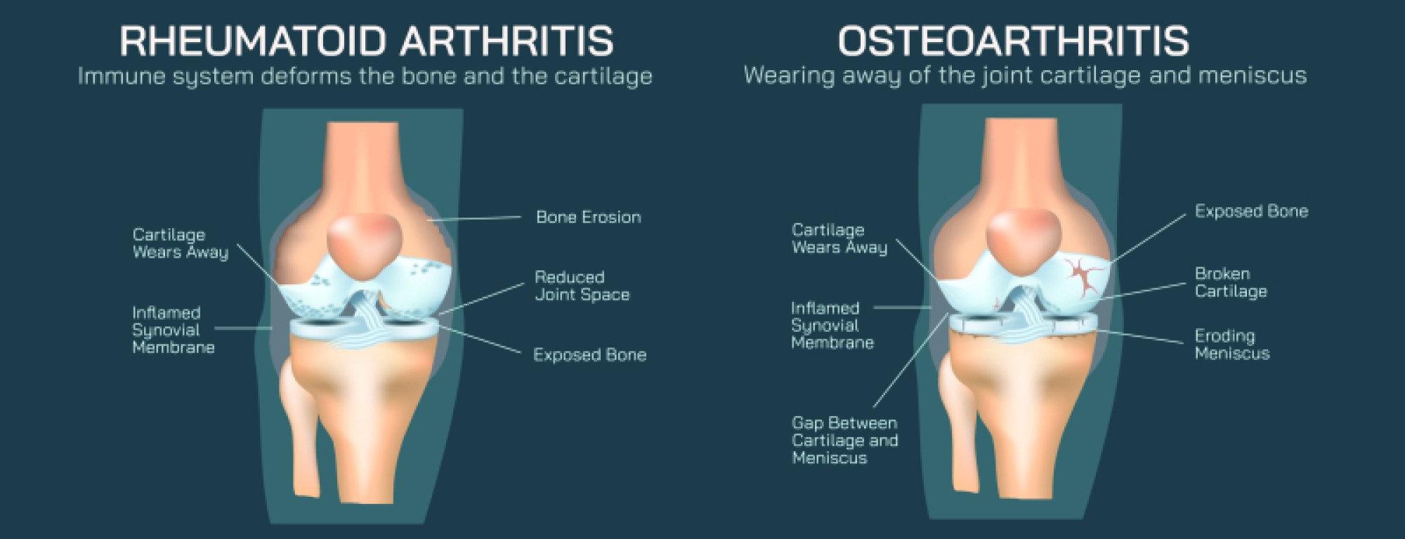
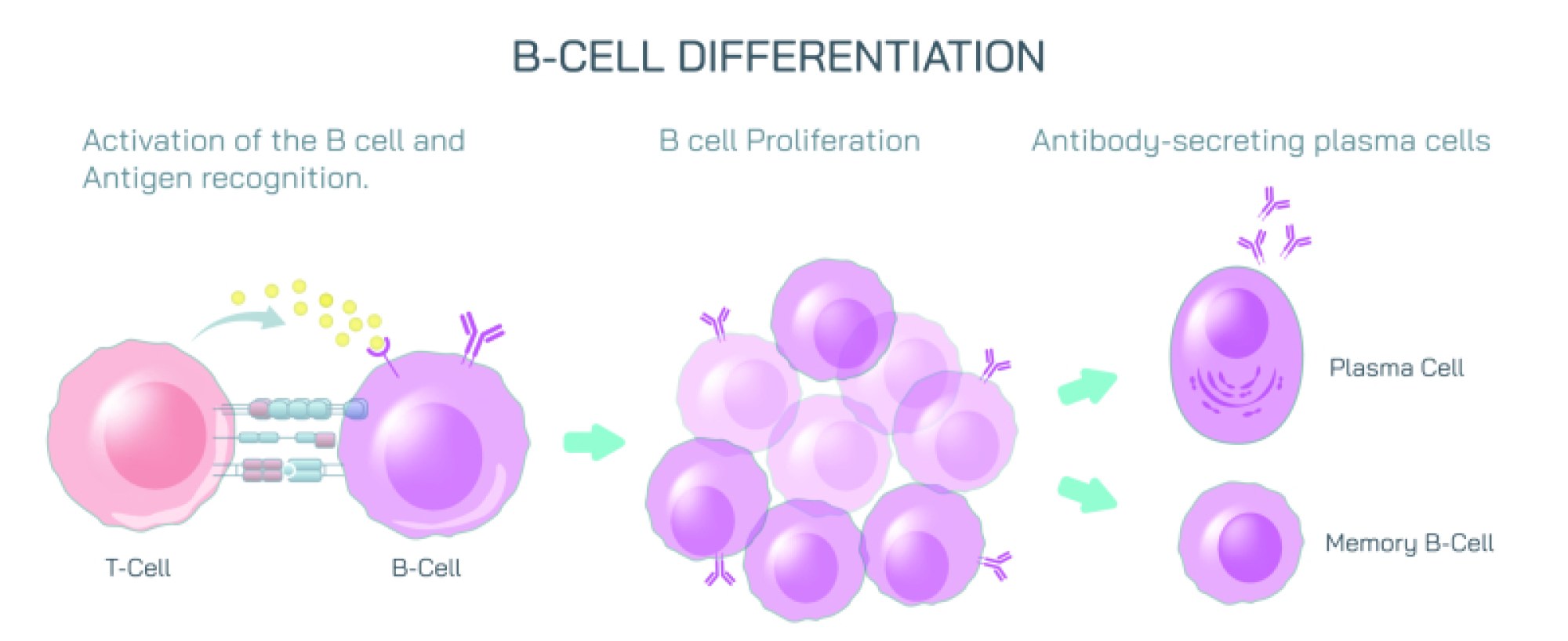
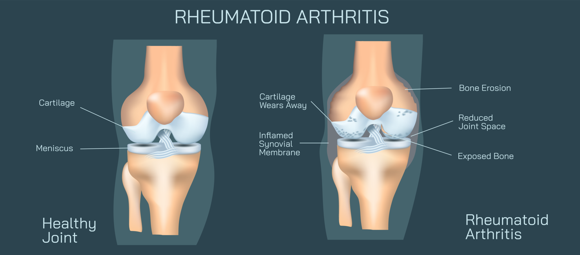
The inflammatory cascade begins in the synovial membrane, which normally appears as a thin, delicate lining supporting joint lubrication. In rheumatoid arthritis, the membrane thickens dramatically due to infiltration of immune cells such as macrophages, T-cells, B-cells, and inflammatory cytokines. The tissue becomes red, swollen, and hyperactive—a transformation often illustrated in medical diagrams by the bright thickening of the membrane and the accumulation of inflammatory fluid within the joint capsule. The inflamed synovial tissue begins producing enzymes that break down cartilage and attack surrounding bone surfaces. In a healthy joint, cartilage provides a smooth protective layer between bones; in a rheumatoid joint, this cartilage gradually erodes as the autoimmune inflammation continues. The loss of cartilage exposes raw bone, making even slight movements painful as the bones grind more directly against each other. Alongside this cartilage destruction comes a pathological growth known as pannus—an aggressive inflammatory tissue that expands into the joint space, damaging cartilage and bone further. In cross-sectional illustrations, this pannus appears as thick invasive tissue creeping along the joint surfaces, symbolizing how the disease transforms natural tissues into destructive agents.
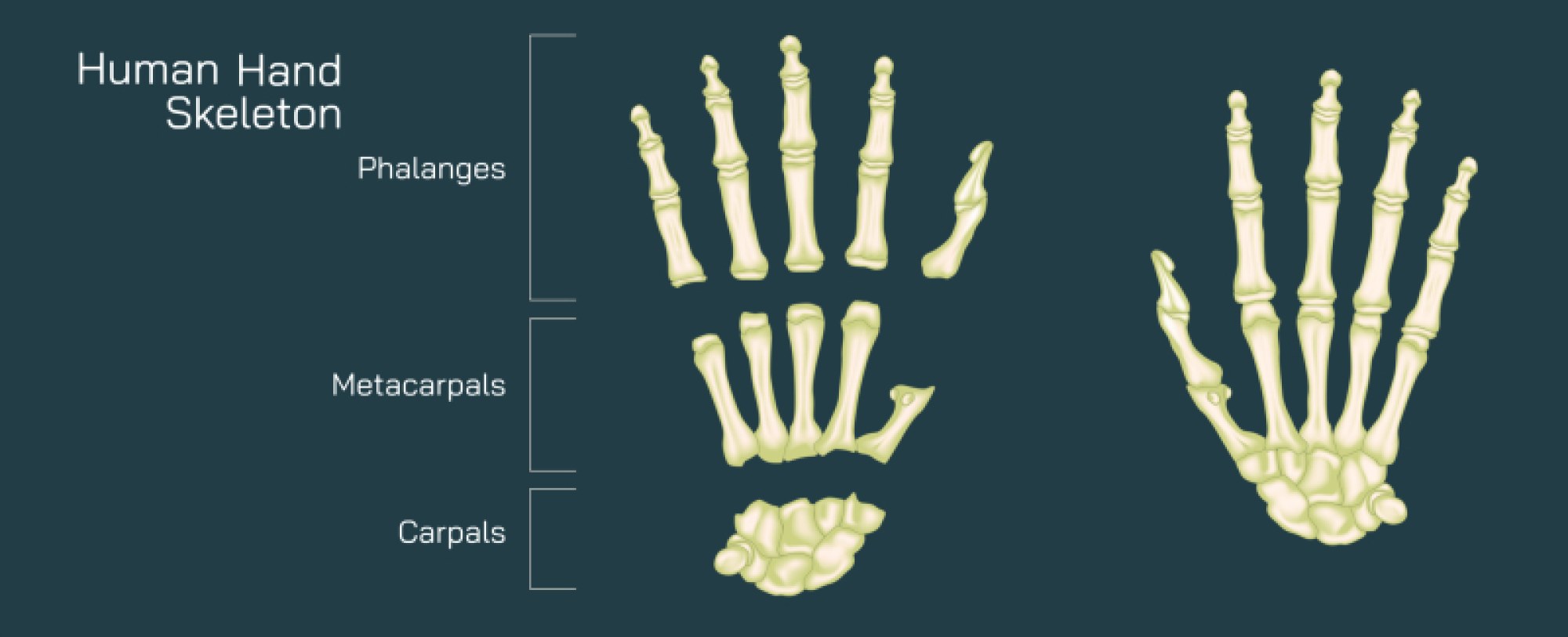
As damage progresses, the effects become visible not only internally but externally through joint deformities and loss of function. The supporting ligaments that stabilize the bones begin to stretch and weaken due to constant inflammation, allowing the joint to lose its alignment. In the fingers and hands, where rheumatoid arthritis is especially common, these changes can result in distinct deformities such as ulnar deviation, swan-neck deformity, and boutonnière deformity. The contrast between the balanced anatomical geometry of a normal joint and the distorted shapes seen in rheumatoid arthritis conveys the dramatic structural changes brought about by chronic inflammation. Muscle wasting around affected joints can also develop as pain and swelling limit normal activity, causing further imbalance between supporting tissues and joint motion. The disease therefore becomes more than localized inflammation; it transforms the biomechanical relationship between bones, cartilage, ligaments, and muscles in a way that reshapes mobility itself.
The immune misidentification that drives rheumatoid arthritis remains an active area of scientific investigation, and an illustration of the disease also reflects this deeper biological struggle. Rheumatoid arthritis is not caused by age-related wear and tear but by autoimmunity—an inappropriate immune response in which the body’s defense system attacks joint tissues as though they were dangerous invaders. Immune cells in the synovium release large amounts of inflammatory molecules such as TNF-alpha, interleukin-1, and interleukin-6, which keep the inflammatory cycle alive rather than allowing it to resolve. These cytokines send chemical signals that recruit even more immune cells to the joint, much like a false alarm that continually calls additional responders to a crisis that does not exist. In diagrams of cell-level disease activity, this process is depicted as a swarm of immune cells congregating around synovial tissue and releasing inflammatory molecules that cause swelling, pain, and tissue destruction. Because the underlying process is systemic rather than localized, rheumatoid arthritis often affects multiple joints symmetrically, and patients may also experience fatigue, anemia, fever, or inflammation in other organs such as the lungs, eyes, and blood vessels. The disease illustrates how autoimmunity can begin in a specific tissue yet eventually echo throughout the body.
The experience of pain in rheumatoid arthritis comes from multiple sources, and anatomical illustrations reveal these layers. Swelling in the joint capsule stretches pain-sensitive nerves in the surrounding tissues. The erosion of cartilage exposes nerve-rich bone surfaces, making even small weight-bearing movements acutely uncomfortable. Inflammation of tendons and bursae adds further signals of pain. The surrounding muscles tighten defensively, creating stiffness that patients commonly describe in the morning or after periods of inactivity. Unlike some forms of arthritis that improve with use, rheumatoid arthritis often remains painful even as movement continues, because the underlying inflammatory problem persists regardless of mechanical changes. The chronic nature of this pain shapes quality of life, sleep patterns, and emotional wellness, reinforcing the idea that rheumatoid arthritis is not only a mechanical problem of the joints but a complex pain disorder linked to immune dysregulation.
Medically illustrated diagrams often compare a healthy joint and a joint affected by rheumatoid arthritis to highlight the differences in structure and fluid balance. In the healthy joint, the synovial cavity appears narrow and calm, cartilage smooth and thick, and bones properly aligned. In the rheumatoid joint, the synovial membrane thickens dramatically, the joint capsule expands with excess synovial fluid, and the cartilage layer becomes visibly eroded. Pannus tissue appears encroaching like a living organism widening its territory, while bone surfaces become irregular and porous as erosion progresses. Ligaments stretch outward rather than holding bones in place, creating visible misalignment that shapes many of the recognizable physical features of rheumatoid arthritis. Such anatomical comparisons play an essential educational role by turning abstract medical descriptions into visual realities that help patients, caregivers, and clinicians understand how subtle changes eventually lead to major functional decline.
Yet the story of rheumatoid arthritis is not solely one of deterioration; it is also one of progress in medical science, empathy, and treatment evolution. Even though the autoimmune process cannot currently be cured, modern therapies aim to interrupt the destructive inflammatory cycle. Disease-modifying antirheumatic drugs and newer biological therapies target specific components of the immune response, such as overactive cytokines or hyperactive immune cells, slowing disease progression, reducing inflammation, and preserving joint structure. Physiotherapy, occupational therapy, and tailored exercise strategies help maintain strength, mobility, and independence. Assistive devices and ergonomic tools protect joints and encourage energy-efficient movement. Illustrations of rheumatoid arthritis today often include depictions of both pathology and pathways of treatment, emphasizing that scientific understanding can change the future course of the disease rather than merely describe its effects.
At its core, rheumatoid arthritis represents a profound example of how structure and function intertwine in human biology. The joint is not simply a hinge of bone and cartilage but a complex living organ where mechanical function depends entirely on the harmony of immune, muscular, structural, and neurological systems. When that harmony is disrupted by autoimmunity, every layer—from the microscopic lining of the synovium to the alignment of the bones—reflects the consequences. A medical illustration of rheumatoid arthritis, therefore, is more than a diagram of swelling and erosion; it is a visual biography of a disease that begins invisibly yet becomes unmistakable as inflammation reshapes anatomy and motion over time. By recognizing the patterns of change within an inflamed joint, patients and clinicians alike deepen their understanding of the disease and reinforce the importance of early diagnosis, consistent treatment, and continuous care. Through this lens, the visual study of rheumatoid arthritis offers not only insight into the mechanics of autoimmunity but also an appreciation of the resilience of the human body and the ongoing scientific pursuit to protect its structure and restore its function.